Non-rigid 2D/3D registration of coronary artery models with live fluoroscopy images

a fluoroscopy and coronary artery technology, applied in the field of methods, can solve the problems of a vast array of minimally invasive procedures, complicating the intervention, and cto crossing using a guide wire is particularly hazardous, and achieves the effect of reducing the uncertainty inherent in 2d interventional images, facilitating the intervention, and solving the problem fast enough

Active Publication Date: 2015-02-03
SIEMENS HEALTHCARE GMBH
View PDF5 Cites 10 Cited by
  • Summary
  • Abstract
  • Description
  • Claims
  • Application Information

AI Technical Summary

Benefits of technology

[0008]Exemplary embodiments of the invention as described herein generally include systems and methods for a 2D / 3D nonrigid registration that can nonrigidly align a preoperative 3D centerline model of the coronary arteries with two or more intraoperative fluoroscopic images to visually augment the interventional images. A registered model according to an embodiment of the invention can be overlaid on top of the interventional angiograms to provide surgical assistance during image-guided chronic total occlusion procedures, thereby reducing the uncertainty inherent in 2D interventional images. A method according to an embodiment of the invention includes two parts: (1) a global transformation model that is calculated to provide an initial rigid or affine alignment, and (2) a fully nonrigid model that is used to compute the final registration. To estimate the global alignment parameters, distance maps are used to measure the discrepancies between the projections of the 3D model and features automatically extracted from the 2D images. In the second part, a fully nonrigid registration method is used to compensate for any local shape discrepancy. A nonrigid registration method according to an embodiment of the invention is based on a variational framework, and uses a simultaneous matching and reconstruction process to compute a nonrigid registration. With a typical run time of less than 3 s on modern hardware, an algorithm according to an embodiment of the invention can be solved fast enough to be used intraoperatively during CTO procedures. In addition, a 2D / 3D registration method according to an embodiment of the invention can also be used to capture the motion of the structure of interest across a sequence of frames.
[0009]According to an aspect of the invention, there is provided a method for non-rigid registration of digital 3D coronary artery models with 2D fluoroscopic images during a cardiac intervention, including providing a digitized 3D centerline representation of a coronary artery tree, where the 3D centerline comprises a set of S segments composed of Qs 3D control points, globally aligning the 3D centerline to at least two 2D fluoroscopic images, and non-rigidly registering the 3D centerline to the at least two 2D fluoroscopic images by minimizing an energy functional that includes a summation of square differences between reconstructed centerline points and registered centerline points, a summation of squared 3D registration vectors, a summation of squared derivative 3D registration vectors, and a myocardial branch energy, where the non-rigid registration of the 3D centerline is represented as a set of 3D translation vectors rs,q that are applied to corresponding centerline points xs,q in a coordinate system of the 3D centerline.
[0014]According to a further aspect of the invention, the method includes providing a time series of F pairs of 2D fluoroscopic images, and globally aligning the 3D centerline to each pair of 2D fluoroscopic images by minimizing the energy functional

Problems solved by technology

During these procedures, the crossing of the CTO using a guide-wire is particularly hazardous since the occlusion blocks the propagation of the contrast agent, and makes the occluded portion of the vessel invisible under fluoroscopy.
In addition, the projective nature of fluoroscopy results in ambiguities in the interpretation of 3D structures, which further complicates the intervention.
Similar challenges arise in a vast array of minimally invasive procedures.
This makes CTA a highly valuable tool at the planning stage, but establishing correspondence between these data and the interventional images can prove challenging.
However, achieving this alignment is a challenge in itself, mainly because finding intermodal correspondence is nontrivial, and also because of the nonlinear aspect of the underlying optimization.
Furthermore, a simple rigid transformation might not suffice to provide a satisfying 2D / 3D registration.
This is, however, a challenging ill-posed inverse task.
The optimization surface in 2D / 3D registration can be highly nonlinear for several reasons: the discretization of the image, the complexity of the structures to be registered, and the use of a rigid or affine transformation model.
As a result, the minimization of the cost function is challenging.
The maximal precision that can potentially be achieved by a registration process is directly linked to the complexity of the transformation model involved.
In general, this is appropriate for rigid structures, such as bones, or to provide an initial alignment of the modalities, but might prove insufficient to account for the shape changes of flexible structures.
Monoplane scenarios have been investigated, but the reported errors are large in the out-of-plane direction.
This lack of accuracy seriously limits confidence in the registration process.

Method used


Image

  • Non-rigid 2D/3D registration of coronary artery models with live fluoroscopy images
  • Non-rigid 2D/3D registration of coronary artery models with live fluoroscopy images
  • Non-rigid 2D/3D registration of coronary artery models with live fluoroscopy images

Examples


Embodiment Construction

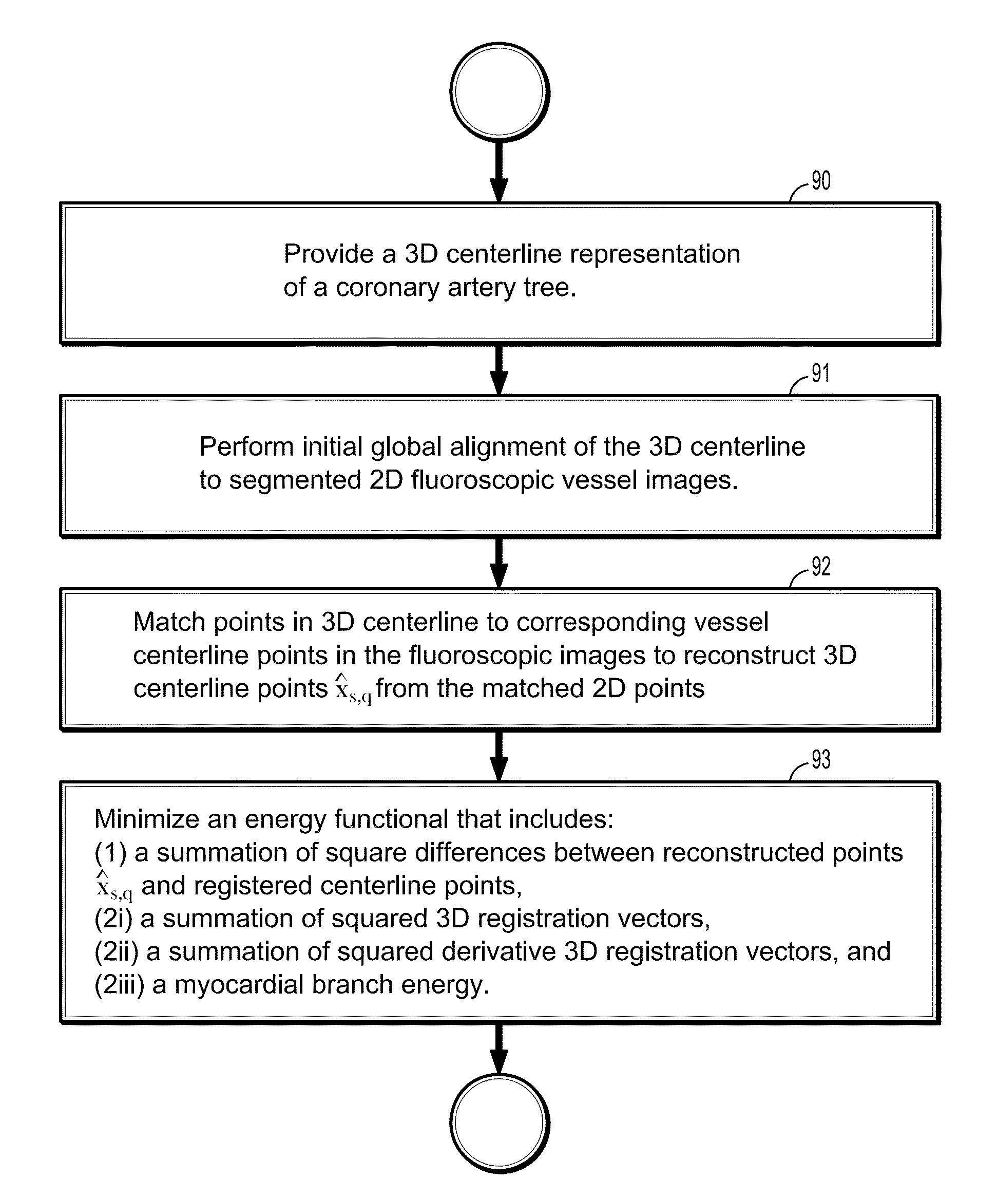
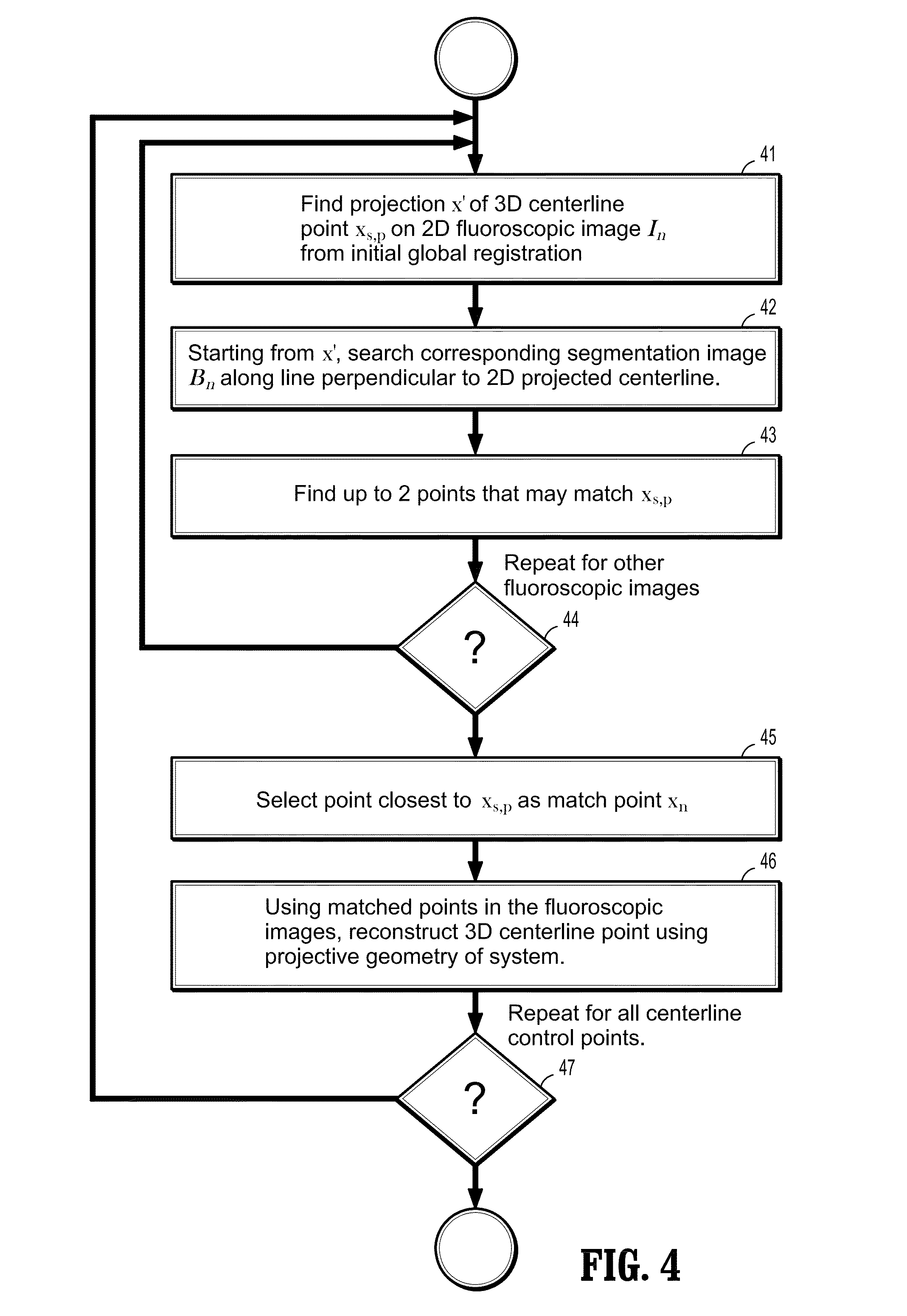
[0044]Exemplary embodiments of the invention as described herein generally include systems and methods for non-rigid 2D / 3D registration of coronary artery models with live fluoroscopy images. Accordingly, while the invention is susceptible to various modifications and alternative forms, specific embodiments thereof are shown by way of example in the drawings and will herein be described in detail. It should be understood, however, that there is no intent to limit the invention to the particular forms disclosed, but on the contrary, the invention is to cover all modifications, equivalents, and alternatives falling within the spirit and scope of the invention.

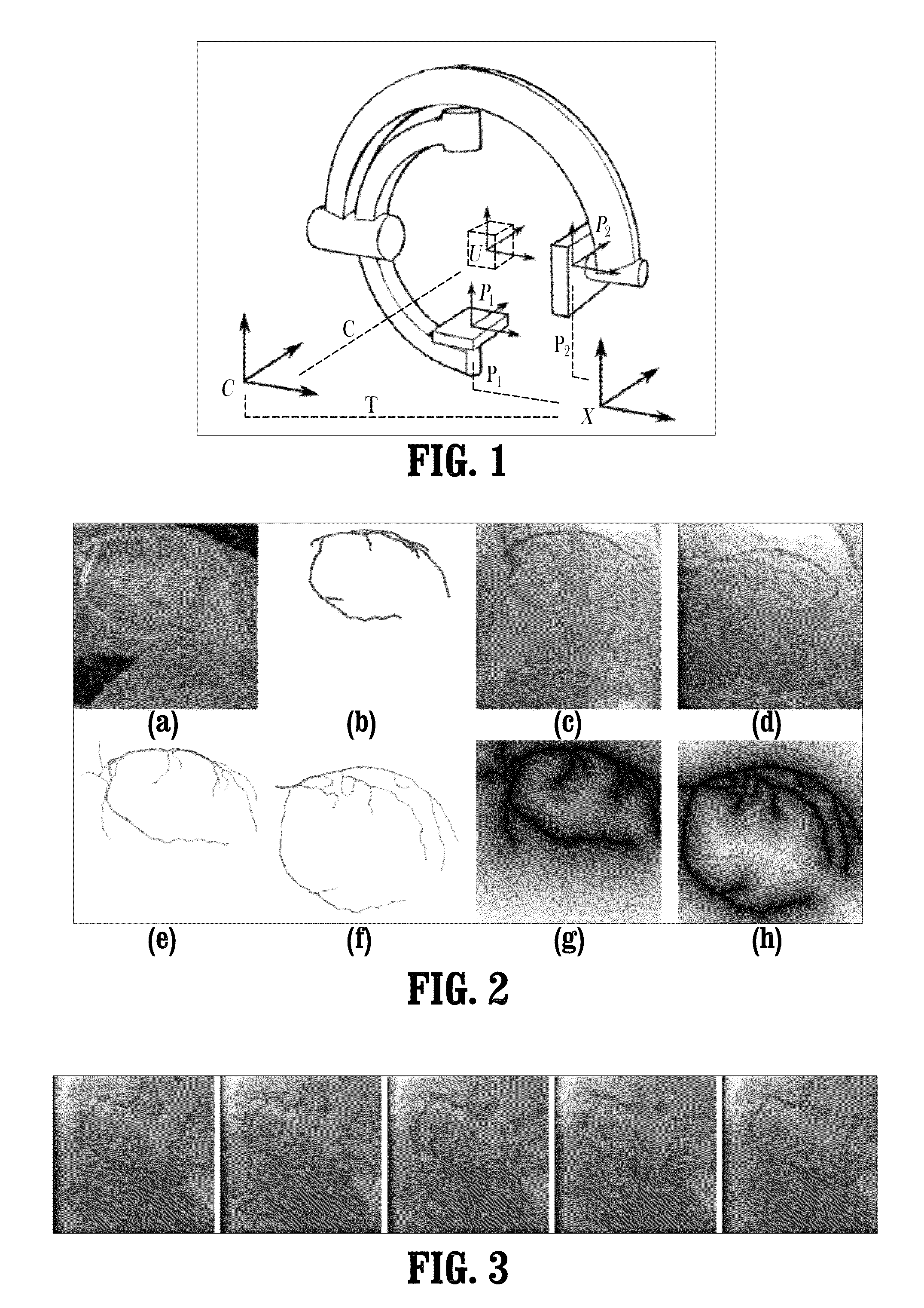
[0045]As used herein, the term “image” refers to multi-dimensional data composed of discrete image elements (e.g., pixels for 2-dimensional images and voxels for 3-dimensional images). The image may be, for example, a medical age of a subject collected by computer tomography, magnetic resonance imaging, ultrasound, or any other m...


PUM


Abstract

A method for non-rigid registration of digital 3D coronary artery models with 2D fluoroscopic images during a cardiac intervention includes providing a digitized 3D centerline representation of a coronary artery tree that comprises a set of S segments composed of QS 3D control points, globally aligning the 3D centerline to at least two 2D fluoroscopic images, and non-rigidly registering the 3D centerline to the at least two 2D fluoroscopic images by minimizing an energy functional that includes a summation of square differences between reconstructed centerline points and registered centerline points, a summation of squared 3D registration vectors, a summation of squared derivative 3D registration vectors, and a myocardial branch energy. The non-rigid registration of the 3D centerline is represented as a set of 3D translation vectors rs,q that are applied to corresponding centerline points xs,q in a coordinate system of the 3D centerline.

Description

CROSS REFERENCE TO RELATED UNITED STATES APPLICATIONS[0001]This application claims priority from “Non-Rigid 2D / 3D Registration of Coronary Artery Models with Live Fluoroscopy for Guidance of Cardiac Interventions”, U.S. Provisional Application No. 61 / 540,134 of Hari Sundar, filed Sep. 28, 2011, the contents of which are herein incorporated by reference in their entirety.TECHNICAL FIELD[0002]This disclosure is directed to methods for non-rigid 2D / 3D registration of anatomical models in digital medical images.DISCUSSION OF THE RELATED ART[0003]X-ray fluoroscopy is the modality of choice for the guidance of many minimally invasive procedures, such as percutaneous coronary interventions (PCI) of chronic total occlusions (CTO). During these procedures, the crossing of the CTO using a guide-wire is particularly hazardous since the occlusion blocks the propagation of the contrast agent, and makes the occluded portion of the vessel invisible under fluoroscopy. In addition, the projective na...

Claims


Application Information

Patent Timeline
Patent Type & Authority Patents(United States)
IPC IPC(8): G06K9/00G06K9/32G06T3/00G06T7/00
CPCG06T3/0068G06T7/0028G06T7/0032G06T3/0081G06T2207/10072G06T2207/10121G06T2207/30101G06T2207/30172G06T7/33G06T7/344
Inventor SUNDAR, HARI
Owner SIEMENS HEALTHCARE GMBH
Why Eureka
Try Eureka