Mediastinoscopy access, sampling, and visualization kit featuring toroidal balloons and exotracheal method of using

a technology of exotracheal and toroidal balloons, which is applied in the field of medical devices, can solve the problems of many problems with conventional devices and approaches to mediastinoscopy, poor visualization, and pain in the neck through the many sensitive muscles and nerves, and achieve the effects of improving access, sampling, and visualization

Inactive Publication Date: 2015-02-10
CHEST INNOVATIONS
View PDF42 Cites 12 Cited by
  • Summary
  • Abstract
  • Description
  • Claims
  • Application Information

AI Technical Summary

Benefits of technology

[0016]The present invention provides a kit for performing a mediastinoscopy that improves access, sampling, and visualization. The various elements of the kit are designed to complement one another and be used together. However, any single element (i.e. the pieces for access only, sampling only, or visualization only) could also be used independently or with other commercially available products for mediastinoscopy or other procedures.
[0018]The toroidal balloons may be used for several purposes. One purpose is to expand a portion of the mediastinal cavity into which light from an endoscope may be directed for improved node visualization prior to grasping. Another purpose is to expand a difficult-to-reach portion of the mediastinal cavity, such as a crevice or narrow interstice between two adjacent structures, for easier access by grasping instruments to a node situated within the interstice. The toroidal balloons can also be used with other regular, non-toroidal balloons that block off an entire portion of the mediastinal cavity to protect sensitive structures or redirect flow, leaving unintended areas undisturbed as the sampling instrument (i.e. MEDIAGOPHER™ sampling instrument as discussed herein) goes after a target node. The atraumatic, flexible surface of an inflatable element such as a balloon provides minimal or no irritation to sensitive structures compared to what a grasping instrument with prongs or a cutting instrument with electrodes and / or blades would do.
[0020]Third, the visualization system comprises a camera that provides a three hundred and sixty degree (360°) view positioned in the “throat” of the MEDIAGOPHER™ sampling instrument. The camera has a broad range of motion. Initially, it is positioned inside the small, narrow, elongated tube upon which the sampling jaw structure is mounted. In this position the camera is proximal to the sampler and the toroidal balloons. The camera can also be advanced to the region just between the sampling jaws for a direct close-up view of the sampling action. The camera can be advanced further beyond the MEDIAGOPHER™ sampling instrument and through more distal expanded toroidal balloons to explore an area before the MEDIAGOPHER™ sampling instrument jaws or other sampling instruments go there. The camera may also be advanced before balloons are advanced and expanded if it is unlikely to cause agitation. This on-site check keeps the sampler on track and confirms that the directions provided by any secondary navigation system (i.e. grid or coordinate system using ultrasound or X-ray data) are accurate. Optionally, communication between transmitters and receivers on the camera and already advanced balloons may be used to set-off expansion of the balloons just before or as the camera passes through them. By using the visualization system to explore a region before advancing other tools the trauma to the patient is minimized. Additionally, misalignment of the patient's body with a secondary navigation system can be quickly detected when the secondary system is instructing for the sampling instruments to move into an area in which there is no abnormal tissue, as seen by the on-site camera. Finally, according to a preferred embodiment, the 360° camera is also capable of doing a U-turn to look back upon the sampling jaws of the MEDIAGOPHER™ sampling instrument from a location distal to the sampling action to watch the biopsy process as it occurs from another perspective. This ability to do a U-turn compensates for any blind spot that may otherwise exist, even with “eyes in the back” and a 360° field of view, at a point where the instrument shaft attaches to the camera (for wiring, etc.).
[0022]The present invention provides a system and method to overcome the shortcomings in the reference art by focusing on the initial access phase of a mediastinoscopy procedure for lymph node sampling. The invention can be complementary or supplementary to existing mediastinoscopy tools by improving target visualization and access prior to the insertion of resecting tools with blades, electrodes, vacuums and compartmentalized collection chambers.

Problems solved by technology

Problems with conventional devices and approaches for mediastinoscopy are numerous.
This leaves a visibly obvious, slow-healing, and painful scar through the many sensitive muscles and nerves in the neck.
Second, visualization is typically poor and even with the assistance of an endoscopic monitor and ultrasound, skilled surgeons have difficulty accessing nodes and assessing whether a node, nodule, or tumor they are about to resect or sever from surrounding tissue is in fact the intended target.
Additionally, operating within dark narrow working spaces increases the risk that the surgeon will inadvertently injure or at least aggravate critical vulnerable structures (including coronary arteries, valves, the heart itself) or puncture a lung in the same general region as the nodes while trying to reach the nodes.
Third, the elongated instruments presently available require reaching out to grasp nodes, cutting, and dangling the severed, potentially abnormal tissue within the cavity prior to removal.
This method risks dropping the sample prior to removal and can cause scattering of malignant particulate material for redistribution in the body amongst healthy tissue.
This system and method however, do not address improving the initial visualization of and access to nodes in crevices or at angles out of the direct trajectory through which an instrument has been inserted.
No patents can be found directly addressing atraumatic mechanical dilation of the mediastinal space.
None of the above patents provide systems, kits, or methods to dilate, easily sample, and improve visualization in the region outside the trachea during a mediastinoscopy procedure.
Further, none of the above patents suggest accessing nodules on the outer trachea, bronchi, mediastinum, or lymph nodes without a neck incision by way of a natural orifice (including the mouth or nose) and natural lumens (including the throat and bronchi).

Method used


Image

  • Mediastinoscopy access, sampling, and visualization kit featuring toroidal balloons and exotracheal method of using
  • Mediastinoscopy access, sampling, and visualization kit featuring toroidal balloons and exotracheal method of using
  • Mediastinoscopy access, sampling, and visualization kit featuring toroidal balloons and exotracheal method of using

Examples


Embodiment Construction

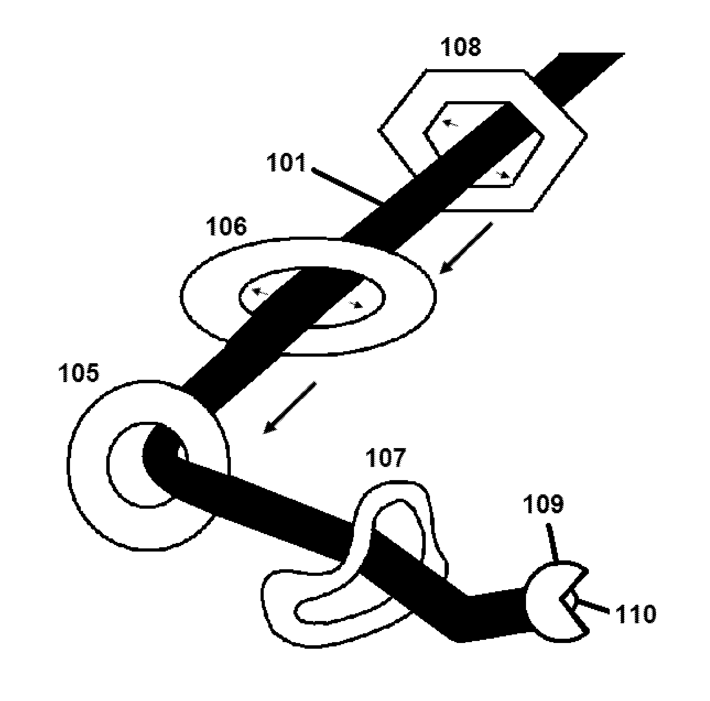
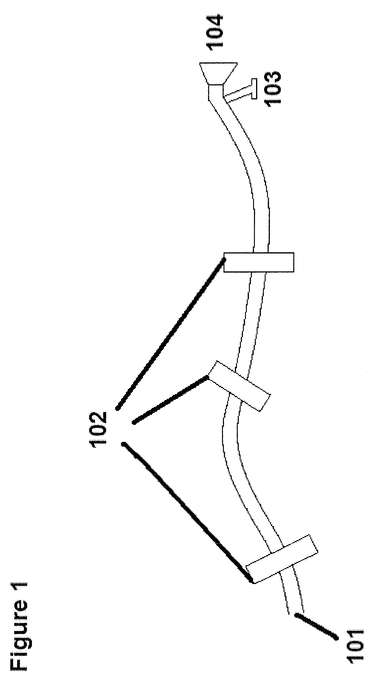
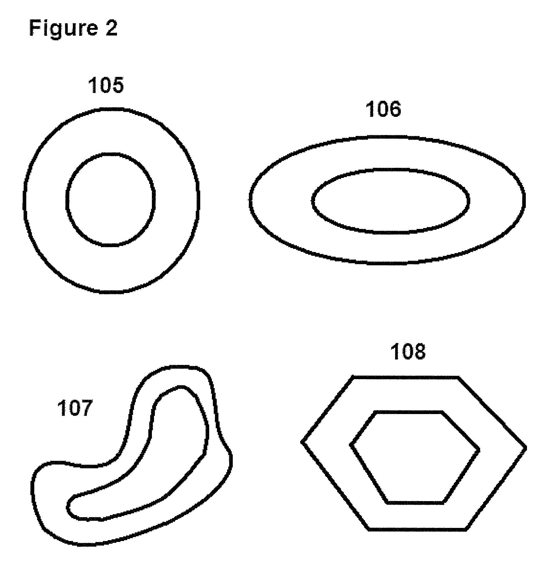
[0030]For the access system the toroidal balloons can have any one of several shapes (including but not limited to donut, elliptical, oblong curved, irregularly curved, and polygonal) and sizes. The shape, size, and material of the balloons may be specially designed or selected depending on the shape, size, and other features of a canal, lumen, or cavity that must be dilated and through which other working instruments will be introduced. Therefore, the toroidal balloons can be chosen based on an individual's anatomy. The size of the toroidal balloons should be specially adapted to fit within the mediastinum including inside the trachea when deflated and outside the trachea when inflated. The shape, size, and material of the balloons can also be tailored to accommodate the working instruments themselves. The material and thickness chosen will influence the flexibility, strength, burst-resistance, maximum pressure, and other properties of the balloon. For example, smaller constricted ...


PUM


Abstract

The present invention provides various systems, a kit, and a method for accessing, sampling within, and visualization of areas within the mediastinal cavity for assisting a surgeon in performing a mediastinoscopy procedure. The access system includes one or more preferably toroidal balloons that can be expanded to dilate and protect the inner walls of a bodily conduit. Instruments pass through hollow spaces within the expanded toroidal balloons. The proximally positioned balloons are expanded first and the unexpanded balloons to be positioned distally are passed through them and subsequently expanded. The sampling system includes an instrument with a rounded head having two or more jaws and a slit therein at the distal end of an elongated tubular body. The visualization system includes a 360° camera that can be positioned from proximal to distal a target site and can also do a U-turn about its axis of extension.

Description

BACKGROUND OF THE INVENTION[0001]1. Field of the Invention[0002]The present invention relates to medical devices designed to assist a physician in performing a mediastinoscopy procedure. More specifically, the invention provides a kit of various systems that work together to improve access, sampling, and visualization during the procedure. Most specifically, the invention relates to inflatable devices for dilating a lumen or cavity in order to prepare a target site, including a mediastinal region, for access by instruments.[0003]2. Description of the Related Art[0004]The term mediastinoscopy refers to an examination of the mediastinum through an incision above and behind the sternum (breastbone) with a suprasternal incision. The mediastinum is the partition separating the right and left thoracic cavities. It is formed from the two inner pleural walls and includes all of the viscera of the thorax except for the lungs. More specifically, the organs in the mediastinal region include th...

Claims


Application Information

Patent Timeline
Patent Type & Authority Patents(United States)
IPC IPC(8): A61M29/00A61M29/02A61M25/10A61B1/00A61B1/313A61B10/06A61B17/00A61B17/02A61B17/22A61B17/29A61B18/14A61B19/00A61B19/04
CPCA61B17/0218A61B1/00082A61B1/313A61B10/06A61B18/1445A61B19/04A61B19/5212A61B2017/00544A61B2017/00557A61B2017/00858A61B2017/22055A61B2017/22068A61B2017/22069A61B2017/2905A61M29/00A61B42/00A61B90/361
Inventor HAMEED, SALMAAN
Owner CHEST INNOVATIONS
Why Eureka
Try Eureka