Increase of myeloid microvesicles in the cerebrospinal fluid as biomarker of microglia/macrophage activation in neurological disorders

a biomarker and cerebrospinal fluid technology, applied in the field of cerebrospinal fluid myeloid microvesicles as biomarkers of microglia/macrophage activation in neurological disorders, can solve the problems of brain context that has never been explored, organelles may be present in vivo at concentrations varying, etc., and achieves easy access, decreases with recovery, and increases with eae severity

a biomarker and cerebrospinal fluid technology, applied in the field of cerebrospinal fluid myeloid microvesicles as biomarkers of microglia/macrophage activation in neurological disorders, can solve the problems of brain context that has never been explored, organelles may be present in vivo at concentrations varying, etc., and achieves easy access, decreases with recovery, and increases with eae severity

US8999655B2Inactive Publication Date: 2015-04-07OSPEDALE SAN RAFFAELE SRL +2

Image

  • Increase of myeloid microvesicles in the cerebrospinal fluid as biomarker of microglia/macrophage activation in neurological disorders
  • Increase of myeloid microvesicles in the cerebrospinal fluid as biomarker of microglia/macrophage activation in neurological disorders
  • Increase of myeloid microvesicles in the cerebrospinal fluid as biomarker of microglia/macrophage activation in neurological disorders

Examples


Embodiment Construction

Materials and Methods

Isolation of CSF MVs

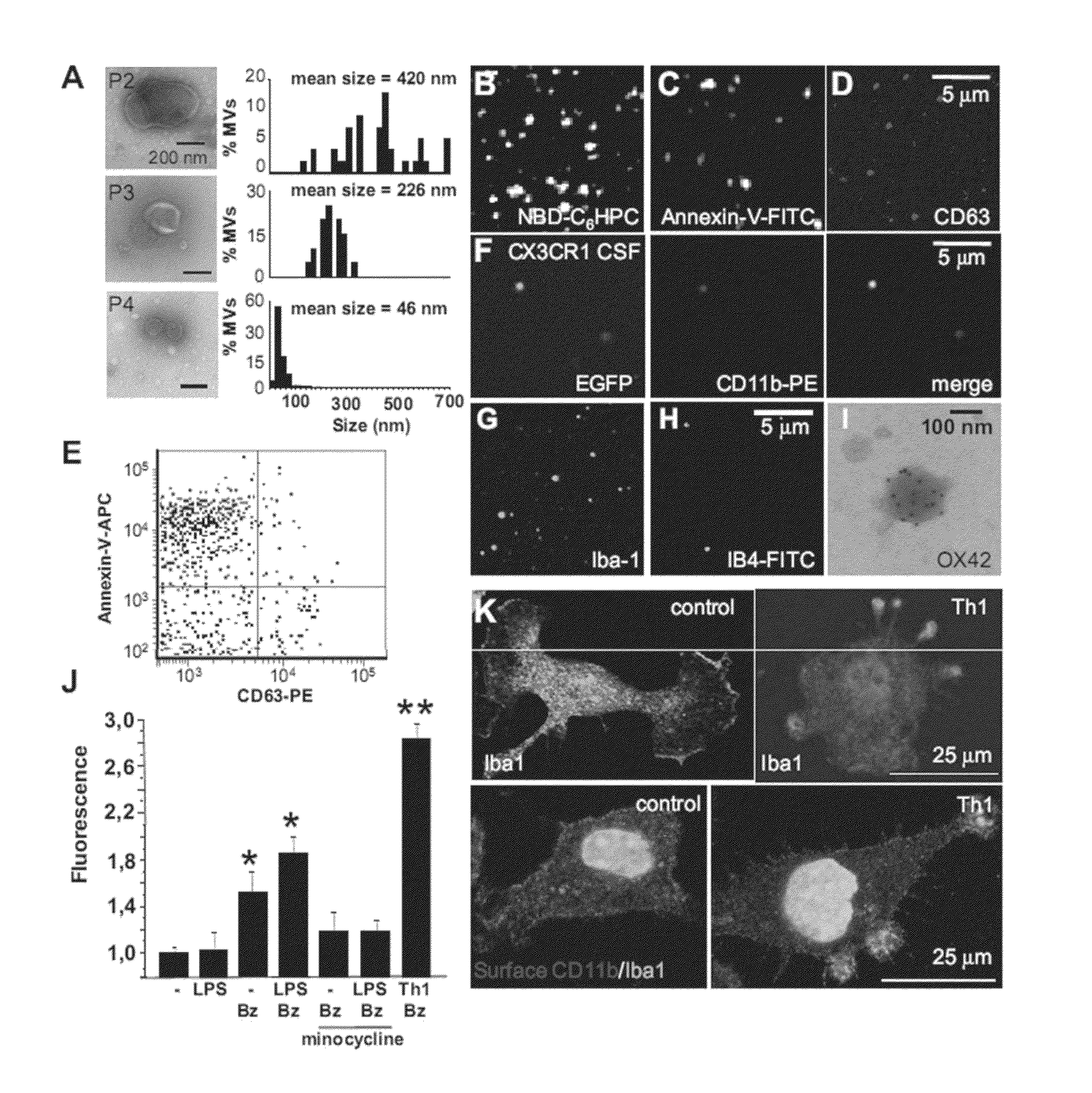
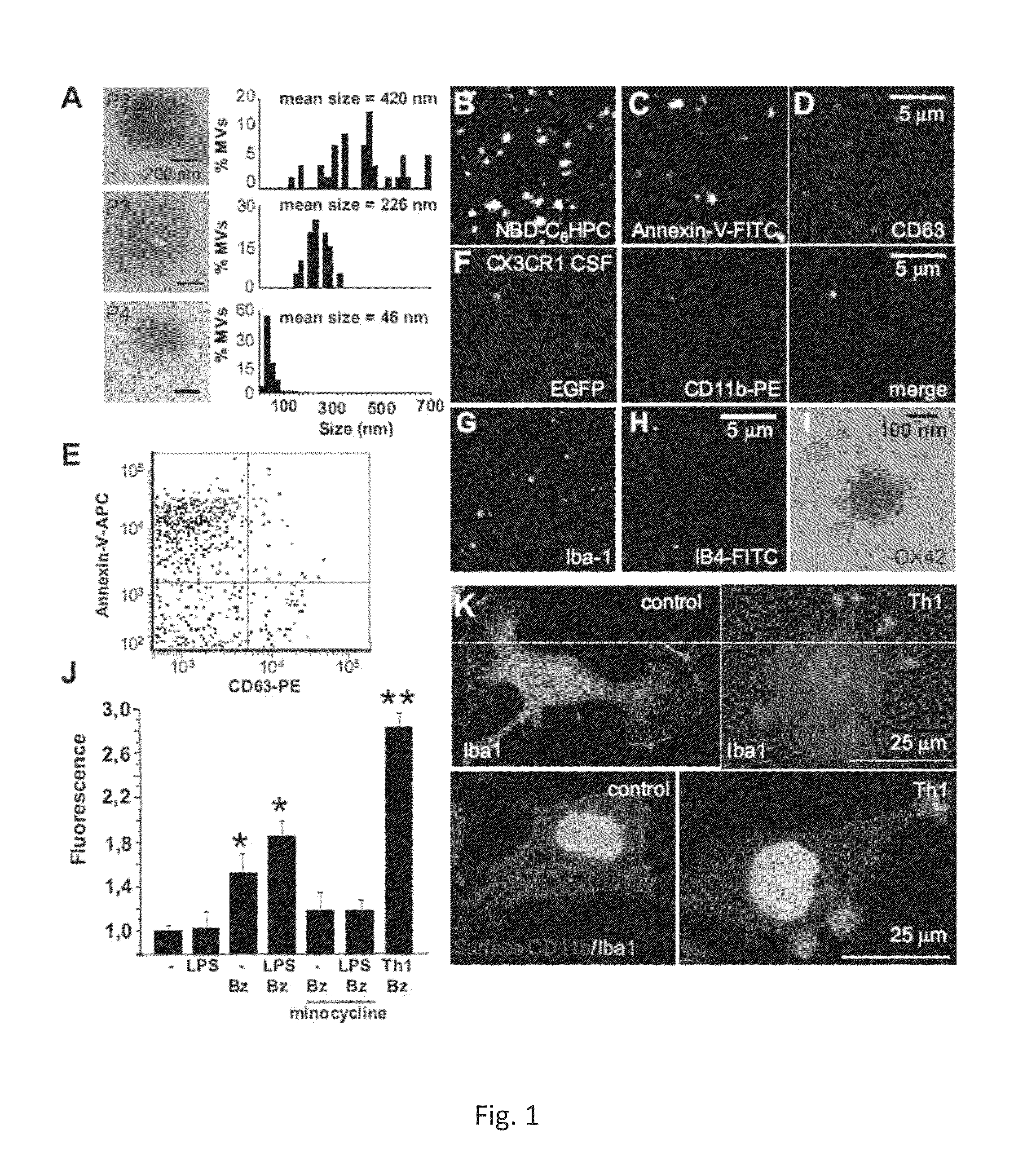
[0048]To collect CSF, rats or mice have been anaesthetized by intraperitoneal injection of 4% Chloral solution and CSF has been sampled from the cisterna magna using a glass capillary (about 10 μl / mouse; about 100 μl / rat) and checked for the absence of blood contamination. CSF pooled from 2-5 rats has been diluted with 0.5-1 ml ice-cold PBS containing protease inhibitors and subjected to differential centrifugation to obtain three vesicles pellets, P2, P3 and P4 pellets, as previously described (Bianco et al., 2009). The resulting pellets were either re-suspended in SDS sample buffer for western blotting, or re-suspended (and fixed when needed) for negative staining electron microscopy or fluorescence microscopy.

Electron Microscopy Negative Staining and Immunogold

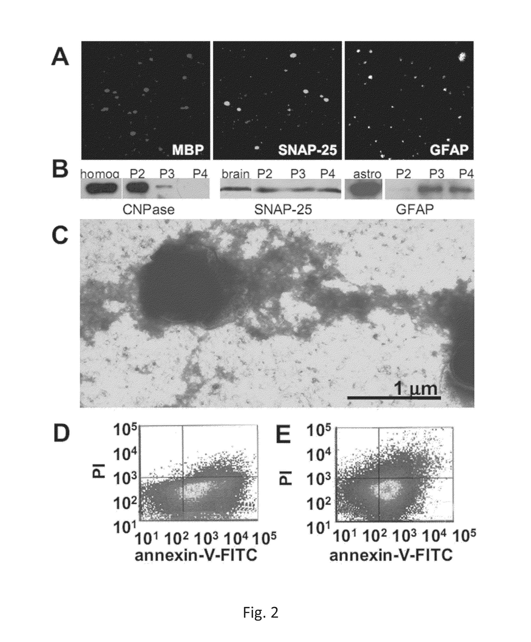
[0049]P2, P3 and P4 MVs or apoptotic bodies from UV irradiated N9 microglial cells were fixed with 4% paraphormaldeyde and adsorbed to 400-mesh formvar / carbon-coated grids; grids wit...


PUM

PropertyMeasurementUnit
sizeaaaaaaaaaa
diameteraaaaaaaaaa
sizeaaaaaaaaaa

Abstract

The present invention relates to a method for the diagnostic and / or prognostic of a neurological disease characterized by an inflammation process in a subject comprising measuring the amount of myeloid derived microvesicles in a cerebrospinal fluid sample obtained from the subject. The invention further relates to a method for predicting and / or monitoring the efficacy of a treatment for a neurological pathology or for monitoring a neurological disease progression.

Description

CROSS-REFERENCE TO RELATED APPLICATION[0001]This application is a 371 of PCT / IB2011 / 050915, filed Mar. 3, 2011, which claims the benefit of U.S. Provisional Application No. 61 / 310,048, field Mar. 3, 2010, the contents of each of which are incorporated herein by reference.TECHNICAL FIELD[0002]The present invention relates to a method for the diagnosis and / or prognosis of a neurological disease characterized by an inflammatory process as well as a method for predicting and / or monitoring the efficacy of a treatment for a neurological pathology. The methods are based on the measurement of the amount of myeloid derived microvesicles in a cerebrospinal fluid sample.BACKGROUND ART[0003]Microvesicles (MVs) of different sub-cellular origin, and released by most cells, have been recently indicated as a novel way of intercellular communication (Al-Nedawi et al., 2009; Cocucci et al., 2009; Ratajczak et al., 2006). MVs include microparticles shed from the plasma membrane (shed MPs), also called...

Claims


Application Information

Patent Timeline
07 Apr 2015
Publication
US8999655B2
IPC
G01N33/50; G01N33/98; G01N23/22; G01N33/68; C12Q1/68; G01N33/566
CPC
G01N33/6896; G01N33/5076; G01N33/5091; G01N2800/28; G01N2800/52; G01N2800/56
Inventors
VERDERIO, CLAUDIA; MATTEOLI, MICHELA