
Plasma membrane biomarkers preferentially expressed in pancreatic beta cells useful in imaging or targeting beta cells

a technology of pancreatic beta cells and plasma membranes, applied in the field of medical devices, can solve the problems of imposing a heavy burden of morbidity and premature mortality, severe long-term complications, end-stage renal failure and neuropathy, etc., and achieves good specificity and selectivity, the effect of assessing the effectiveness

Active Publication Date: 2013-04-23
UNIV LIBRE DE BRUXELIES +2
View PDF3 Cites 2 Cited by
  • Summary
  • Abstract
  • Description
  • Claims
  • Application Information

AI Technical Summary

Benefits of technology

The present invention is about a method to determine the amount of beta cells (a type of pancreatic cell) in diabetes patients. This is important because there are currently no other ways to measure beta cell mass. The method involves using specific biomarkers that are enriched in beta cells and can be labeled with tracers for imaging. These biomarkers are not induced by inflammation and are not targeted by infiltrating immune cells. This approach allows for non-invasive imaging of islet grafts and can help in detecting and managing diabetes. The biomarkers can also be used to better understand the disease's progression and to assess the effectiveness of therapies.

Problems solved by technology

Diabetes causes severe long-term complications and psychosocial problems, imposing a heavy burden of morbidity and premature mortality.
Diabetes is the leading cause of blindness and visual disability, limb amputation, end-stage renal failure and neuropathy.
Because T2D is increasingly prevalent and developing earlier in life, increasing the duration of the disease by decades, many more people are developing severe diabetic complications, and suffering decreases their life quality and expectancy.
Diabetes incurs many costs, and is particularly expensive because it is life-long and causes major medical problems.
These costs include direct costs from physician and nursing services, hospital services, laboratory services, drugs, education and training of the patients and indirect costs from time lost from work, chronic nursing and general socio-economic support, early retirement, protracted morbidity and premature mortality.
Overall costs of diabetes mellitus, comprising both direct and indirect costs, have been calculated in various countries and are enormous.
Diabetes develops when the body doesn't make enough insulin, cannot use insulin properly, or both, causing glucose to build up in the blood.
Over time, insulin production declines as well, so many people with T2D eventually need to take insulin.
Patients with hyperinsulinemia are at high risk of developing seizures, mental retardation, and permanent brain damage.
Since glucose is the primary substrate used by the CNS, unrecognized or poorly controlled hypoglycemia may lead to persistent severe neurologic damage.
The lack of a cellular model for studying the beta cells, and also the lack of reliable and effective means of cell sorting suitable for this type of cells hinder the study of beta cell functioning and therefore the development of novel methods of treatment of type I and II diabetes.
MRI has the best spatial resolution but its major problem is the ex-vivo labeling procedure and its semi-quantitative nature.
This technique is, however, only usable for transplantation since it relies on ex vivo uptake of the marker by islet cells and can not be used for in vivo imaging of beta cells in the pancreas.
On the other hand, these techniques have a lower spatial resolution as compared to MRI.
This results in dilution of tracer and high backgrounds, making it currently impossible to quantify the beta cells which are scattered over the pancreas in tiny islets (100-300 μm diameter) constituting only 1-2% of the total pancreas mass.
It was recently shown, however, that total eradication of beta cells still resulted in VMAT2 binding, showing that DTBZ is not a good biomarker for imaging pancreatic beta cell mass (Kung et al 2007).
Recently SLC30A8 was identified as an autoantigen and the target of autoantibodies in type 1 diabetes (Wenzlau J M et al., 2007) and it is therefore not useful for beta cell detection.
Due to its very low expression levels this candidate is not useful for beta cell detection.
However, this candidate was too low expressed to be of use in beta cell detection.
This receptor, however, is expressed both in islet beta cells and in alpha cells (Flodgren E et al 2007), hampering its potential as a good beta cell biomarker.
Glibenclamide and fluorodithizone are not specific enough to attain the robust signal to background ratio needed for quantification of beta cell mass via PET imaging.
Unfortunately, all the compounds mentioned above result in too high background levels and non-specific binding to various other intra-abdominal tissues such as kidney and liver.
Unfortunately, complete eradication of beta cells reduced the pancreatic uptake of DTBZ by only 30-40% showing that the compound lacks sufficient specificity for the beta cells (Kung et al 2007) to enable its use to assess beta cell mass.
Sulfatide, however, is also expressed in islet cell innervating Schwann cells and other neural tissues, which may hamper its use in beta cell imaging.
Consequently, there is a lack of specific and reliable markers for the beta cells of pancreatic islets of Langerhans and do not allow reliable beta cell mass quantification.

Method used


Image

  • Plasma membrane biomarkers preferentially expressed in pancreatic beta cells useful in imaging or targeting beta cells
  • Plasma membrane biomarkers preferentially expressed in pancreatic beta cells useful in imaging or targeting beta cells
  • Plasma membrane biomarkers preferentially expressed in pancreatic beta cells useful in imaging or targeting beta cells

Examples


example 1

Selection Strategy for the Identification of Beta Cell Specific Plasma Membrane Proteins

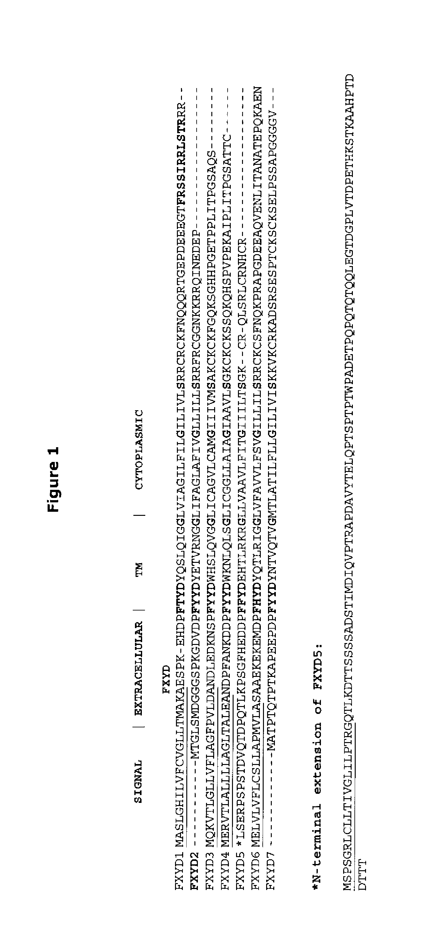
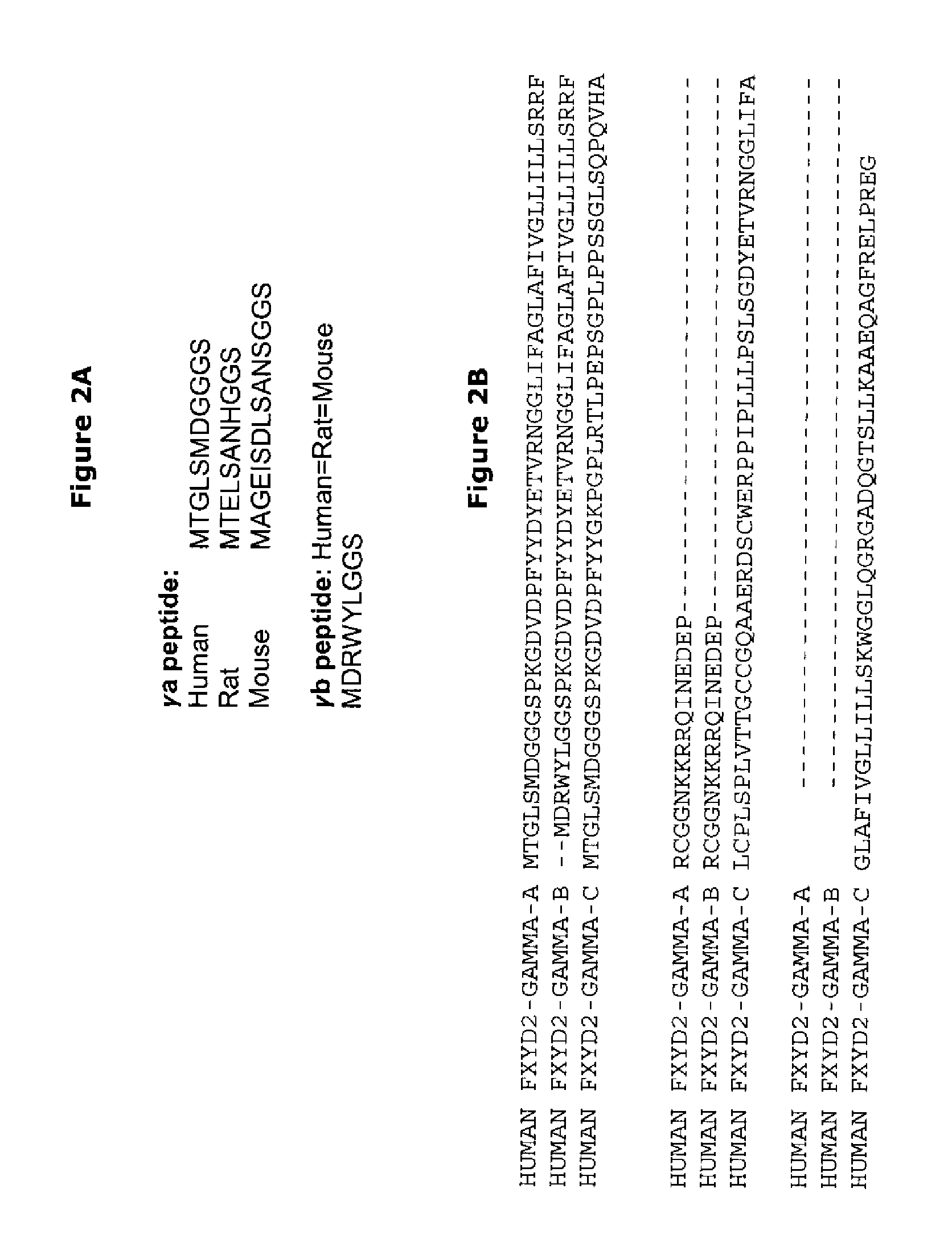
[0178]The strategy used in these examples is depicted in FIGS. 1 and 2. The pancreas is composed of 1-2% of tiny endocrine islets of Langerhans which are scattered in the exocrine tissue. The exocrine pancreas consists of acinar cells and a network of ducts. Islets contain insulin secreting beta cells and non beta cells, e.g. the glucagon secreting alpha cells, pancreatic polypeptide producing PP cells and somatostatin secreting delta cells. Pancreatic beta cells are selectively destroyed by an autoimmune assault in T1D, and there is also evidence that beta cell loss is present in long term T2D, albeit at a lower level than in T1D. Gene expression data on pancreatic islets and on pancreatic beta cells have been obtained in the last 8 years. The methods used are microarray analysis and massive parallel signature sequencing (a procedure that detects gene expression based on sequencing of signatures...

example 2

Validation of the Islet Specific Expression of the Selected Biomarkers

[0181]A. Marker FXYD2:

[0182]MPSS data obtained in human islets compared to expression in 32 human tissues derived from the LICR MPSS dataset; only expression levels above 5 tpm are shown.

[0183]A.1 Introduction:

[0184]The gamma-subunit of the Na, K-ATPase (FXYD2, ATP1G1, HOMG2, MGC12372) is a single-span membrane protein that regulates the Na, K-ATPase by modifying its kinetic properties. The gamma-subunit belongs to a family of 7 FXYD proteins which are tissue specifically expressed (FIG. 1 after Cornelius F et al, 2003). FXYD2 is abundantly expressed in the proximal and distal tubules and in the medullary thick ascending limb of Henle of the kidney (Arystarkhova E et al 2002, Pihakaski-Maunsbach K et al 2006). Two splice variants (gamma-a (SEQ ID NO: 6) and gamma-b SEQ ID NO:7) have been detected in rat and human (Arystarkhova E et al 2005) while the third splice variant gamma-c has been characterized only in mous...

example 3

Confirmation of a Marked Decrease in FXYD Expression in the Pancreas of Two Type 1 Diabetic Patients

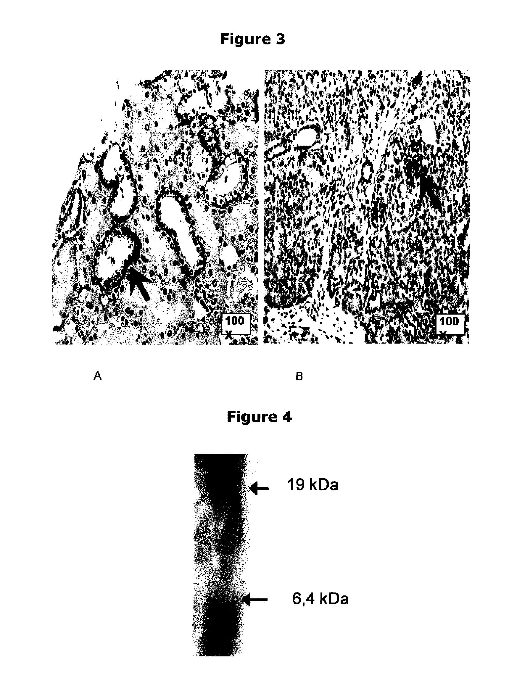
[0318]Expression of FXYD2 gamma-a is drastically decreased in islets from type 1 diabetes patients (CO and TELF; these codes identify two patients, deceased respectively 3 days and 5 years after the diagnosis of type 1 diabetes) compared to normal pancreas (CTRL). Consecutive sections of 3 μm were taken and stained with SPY393 antibody, anti-insulin antibody or anti-glucagon antibody. In the TELF pancreas, beta cells can not be detected based on insulin staining; this was correlated with disappearance of FXYD2 staining. In the CO pancreas, beta cells can not be detected based on insulin staining but a very faint staining of FXYD2 remained (cf. FIG. 12, magnification 1000×).

[0319]FIG. 13 shows the quantification of the FXYD2 gamma-a in islets from type 1 diabetes patients: 20 Langerhans islets / case were quantified; % of stained tissue area (Labelling Index—LI) and mean staining intensi...


PUM

PropertyMeasurementUnit
diameteraaaaaaaaaa
timeaaaaaaaaaa
beta-cell massaaaaaaaaaa

Abstract

The present invention is directed to the identification of a biomarker specifically located in the plasma membrane of pancreatic beta cells. It was selected by a Systems Biology approach on Massively Parallel Signal Sequencing datasets obtained in human islets and Affymetrix microarray datasets on human islets, purified rat primary beta and non beta cells and insulinoma cells. Based on a set of specific features the biomarker is a unique candidate for imaging and targeting strategies to study the pancreatic beta cell mass in health and disease (T1 D, T2D, pancreatic cancers, obesity, islet transplantation, beta cell regeneration). The five specific features of the selected biomarkers are: 1) Preferentially expressed in pancreatic islets as compared to surrounding tissues; 2) Higher expression in pancreatic beta cells than in pancreatic alpha cells or than in other islet non-beta cells; 3) Expression levels in pancreatic beta cells are higher or comparable to glucokinase which is an enzyme specifically expressed in the pancreatic beta cell; 4) Located in the membrane and as such targetable with antibodies, peptides or small molecules which allows imaging, targeting and immunohistochemistry; and 5) Expression is not induced during the process of inflammation of the beta cell mass and the protein is not enriched in T-cells and dendritic cells or in other cells participating in the inflammation process.

Description

CROSS-REFERENCE TO RELATED PATENT APPLICATION(S)[0001]This patent application is the national phase entry of PCT / EP2009 / 051721, filed Feb. 13, 2009, and published, in English, as PCT International Publication WO 2009 / 101181 A2, on Aug. 20, 2009, and claims priority under Article 8 of the Patent Cooperation Treaty and under 35 U.S.C. §119(e) to U.S. provisional patent application No. 61 / 066,068, filed Feb. 14, 2008.STATEMENT OF GOVERNMENT INTEREST[0002]This work was supported by the US Juvenile Diabetes Research Foundation Grant JDRF-4-2001-43. The Juvenile Diabetes Research Foundation may have certain rights in this invention.FIELD OF THE INVENTION[0003]The present invention is directed to the medical field, in particular to the imaging and quantification of pancreatic beta cell mass, targeting or visualization of pancreatic beta cells for pathology and / or diagnosis of diabetic disorders, follow up of islet transplantation and to purification strategies of pancreatic beta cells.BACK...

Claims


Application Information

Patent Timeline
Patent Type & Authority Patents(United States)
IPC IPC(8): A61K49/00
CPCC07K14/705G01N33/573G01N33/57438G01N2800/042
Inventor EIZIRIK, DECIO L.FLAMEZ, DAISYSALMON, ISABELLEBERTON, ALIXROLAND, ISABELLEMARECHAL, DANIELBECKERS, MARIE-CLAIREHOOD, LEROYGOODMAN, NATHANKUTLU, BURAK
Owner UNIV LIBRE DE BRUXELIES
Learn More