Method to reduce loss of cardiac function following ischemia/reperfusion
a technology of ischemia and ischemia, applied in the field of pharmaceutical compositions, can solve the problems of increasing the risk of cardiac dysfunction, affecting the quality of life, so as to confirm the effect and function of mk, and prevent apoptosis of cardiomyocytes
- Summary
- Abstract
- Description
- Claims
- Application Information
AI Technical Summary
Benefits of technology
Problems solved by technology
Method used
Image
Examples
example 1
Expression Pattern of MK Protein in Wild-Type Mouse Heart
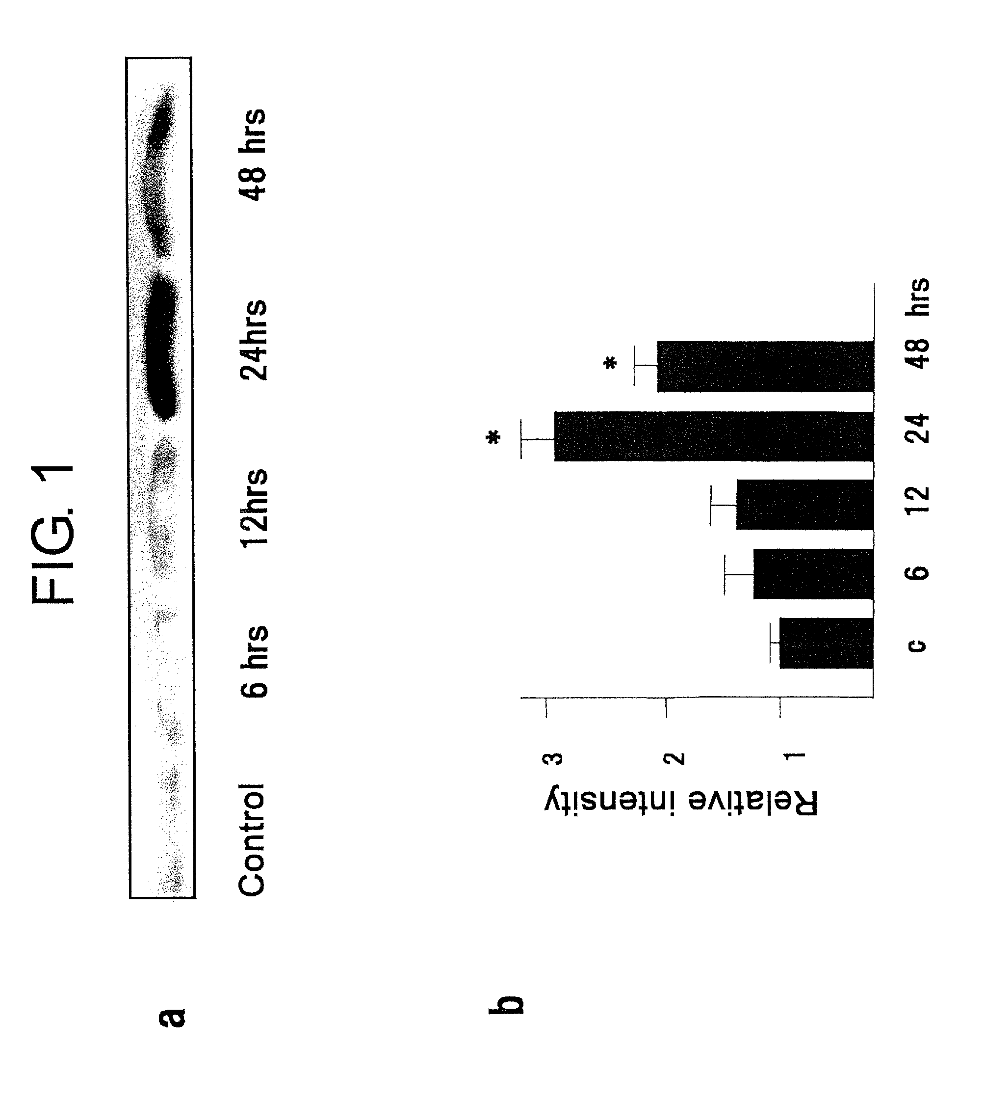
[0051]The time course of MK expression in wild-type mouse hearts after ischemia / reperfusion is shown in FIG. 1. FIG. 1 (a) is an electropherogram obtained by separating 10 mg of the protein extracted from Mdk+ / + mouse hearts through 15% SDS-PAGE and then performing Western blotting; and (b) is a graph showing the intensities of the obtained bands which were quantified with a densitometry. This shows that MK expression in the control is weak, increases with time after ischemia / reperfusion (I / R), and reaches a maximum at 24 hours after I / R, and the increased expression continues up to 48 hours after I / R.
[0052]MK localization was analyzed immunohistochemically in the control cardiac sections of Mdk+ / + mouse hearts and the cardiac sections 24 hours after ischemia / reperfusion (micrographs not shown). In the control cardiac sections, MK protein appeared spread out and pale, but in the cardiac sections 24 hours after ischemia / reperfu...
example 2
Comparison of Myocardial Injuries After Ischemia / Reperfusion in Wild-Type Mice and in Mdk− / − Mice
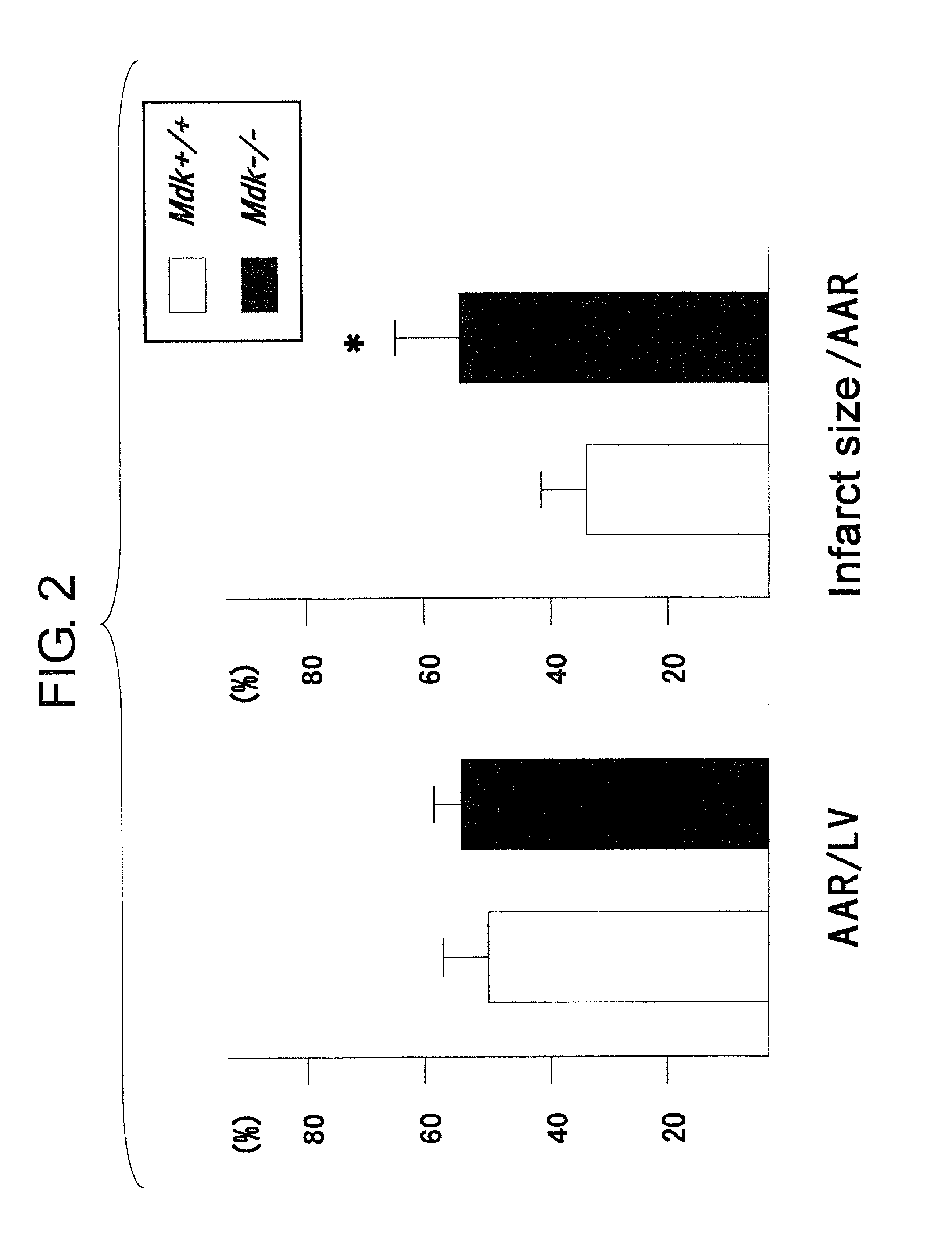
[0053]Myocardial injuries after ischemia / reperfusion were compared using the acute stage Mdk+ / + mouse and Mdk− / − mouse models. In the left ventricular sections, tissues that turned blue by Evans blue staining were identified as areas not at risk, and regions in areas at risk that turned red by TTC staining were identified as tissues that were still alive (micrographs not shown). The regions that were not stained by either Evans blue or by TTC appeared whitish, and this was identified as the infarct region.
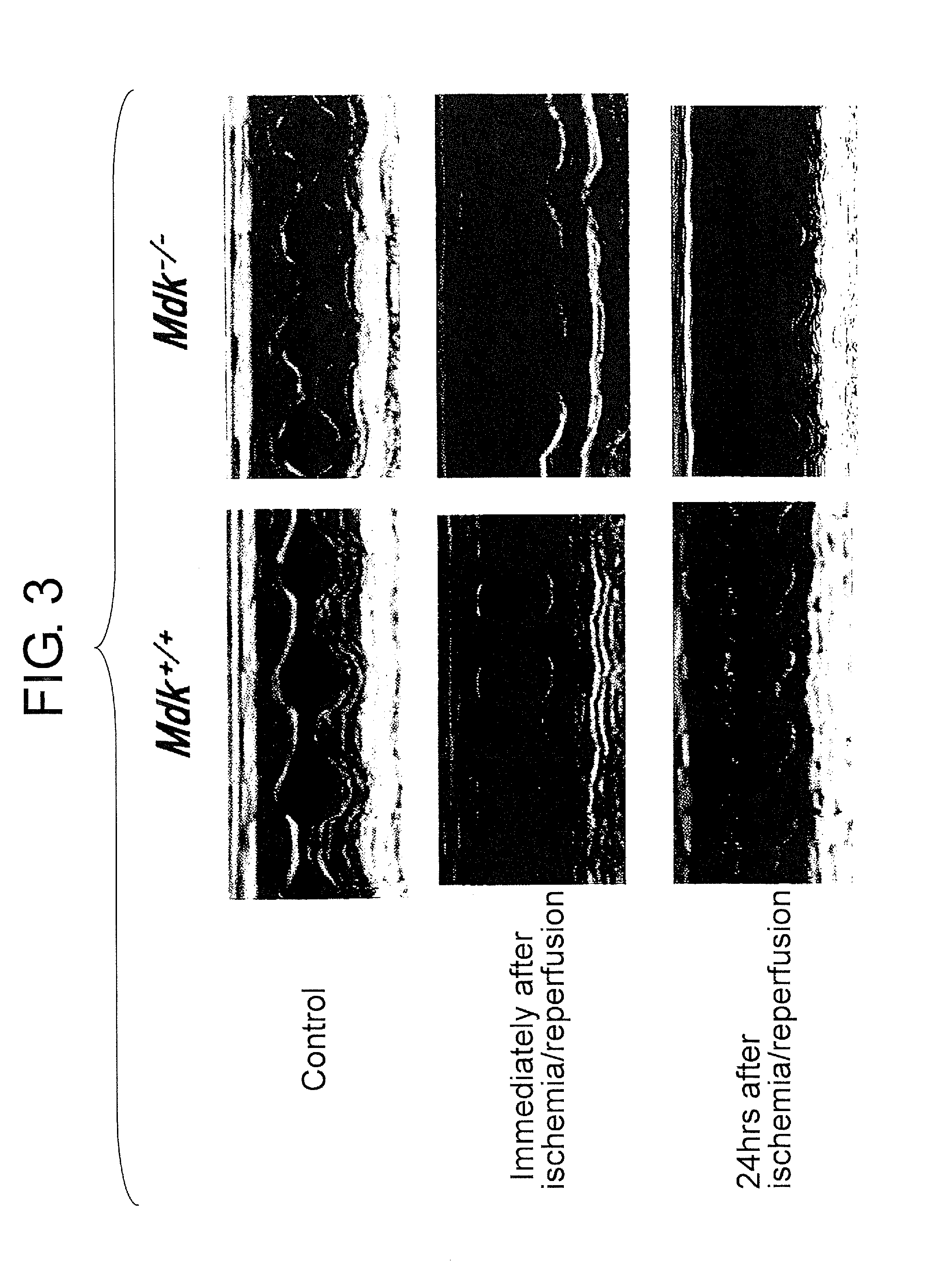
[0054]In FIG. 2, the left graph shows percentages of the infarct areas of the left ventricle (LV) relative to the ischemic left ventricular areas at risk (AAR); and the right graph shows percentages of the infarct size (the white necrotic region) in the AAR. Comparison of Mdk+ / + mice and Mdk− / − mice showed that although areas at risk (AAR) for both mice are similar, the infarct size in ...
example 3
Evaluation of Cardioprotection by MK Protein (In Vitro Experiment)
[0059]Since apoptotic cell death of cultured cardiomyocytes is caused by hypoxia / reoxygenation (H / R), this method was used to evaluate the cardioprotective effect of MK. The cardioprotective effects of the MK protein on cultured cardiomyocytes subjected to H / R treatment are shown in FIGS. 7 and 8. FIG. 7 (a) shows the quantitative DNA fragmentation data determined by ELISA, and (b) shows the number of TUNEL-positive cells relative to the number of nuclei. FIG. 8 (top) shows the changes in Bcl-2 expression level in cardiomyocytes detected by Western blotting. Lane 1: the control, lane 2: the cells subjected to H / R treatment (without MK addition), and lane 3: the cells subjected to H / R treatment (with 100 ng / ml of MK protein).
[0060]After H / R treatment, the amount of DNA fragmentation determined by ELISA and the number of TUNEL-positive cells were both decreased significantly in the presence of 100 ng / ml of MK (FIG. 7). ...
PUM
| Property | Measurement | Unit |
|---|---|---|
| thick | aaaaa | aaaaa |
| left ventricular wall thickness | aaaaa | aaaaa |
| concentrations | aaaaa | aaaaa |
Abstract
Description
Claims
Application Information
 - R&D
- Intellectual Property
- Life Sciences
- Materials
- Tech Scout
- Unparalleled Data Quality
- Higher Quality Content
- 60% Fewer Hallucinations
Browse by: Latest US Patents, China's latest patents, Technical Efficacy Thesaurus, Application Domain, Technology Topic, Popular Technical Reports.
© 2025 PatSnap. All rights reserved.Legal|Privacy policy|Modern Slavery Act Transparency Statement|Sitemap|About US| Contact US: help@patsnap.com


