Method for detecting esophageal carcinoma and agent for suppressing esophageal carcinoma
a technology for esophageal carcinoma and agents, applied in the field of detecting cancer, can solve the problems of extremely poor prognosis of patients expressing smyd2 at high levels, and achieve the effect of suppressing the growth of esophageal carcinoma
- Summary
- Abstract
- Description
- Claims
- Application Information
AI Technical Summary
Benefits of technology
Problems solved by technology
Method used
Image
Examples
example 1
Amplicon Mapping for the 1q32-1q41 Gene Region in ESCC Cell Lines
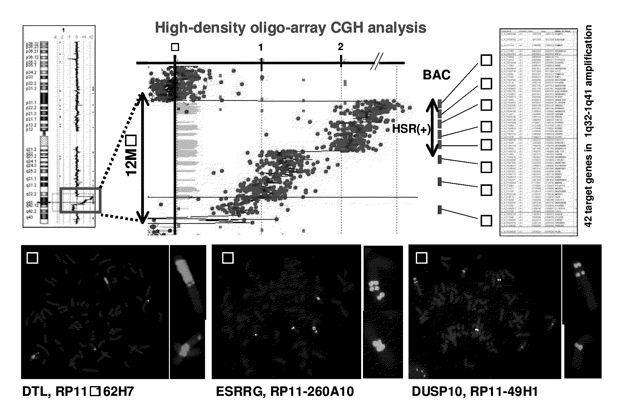
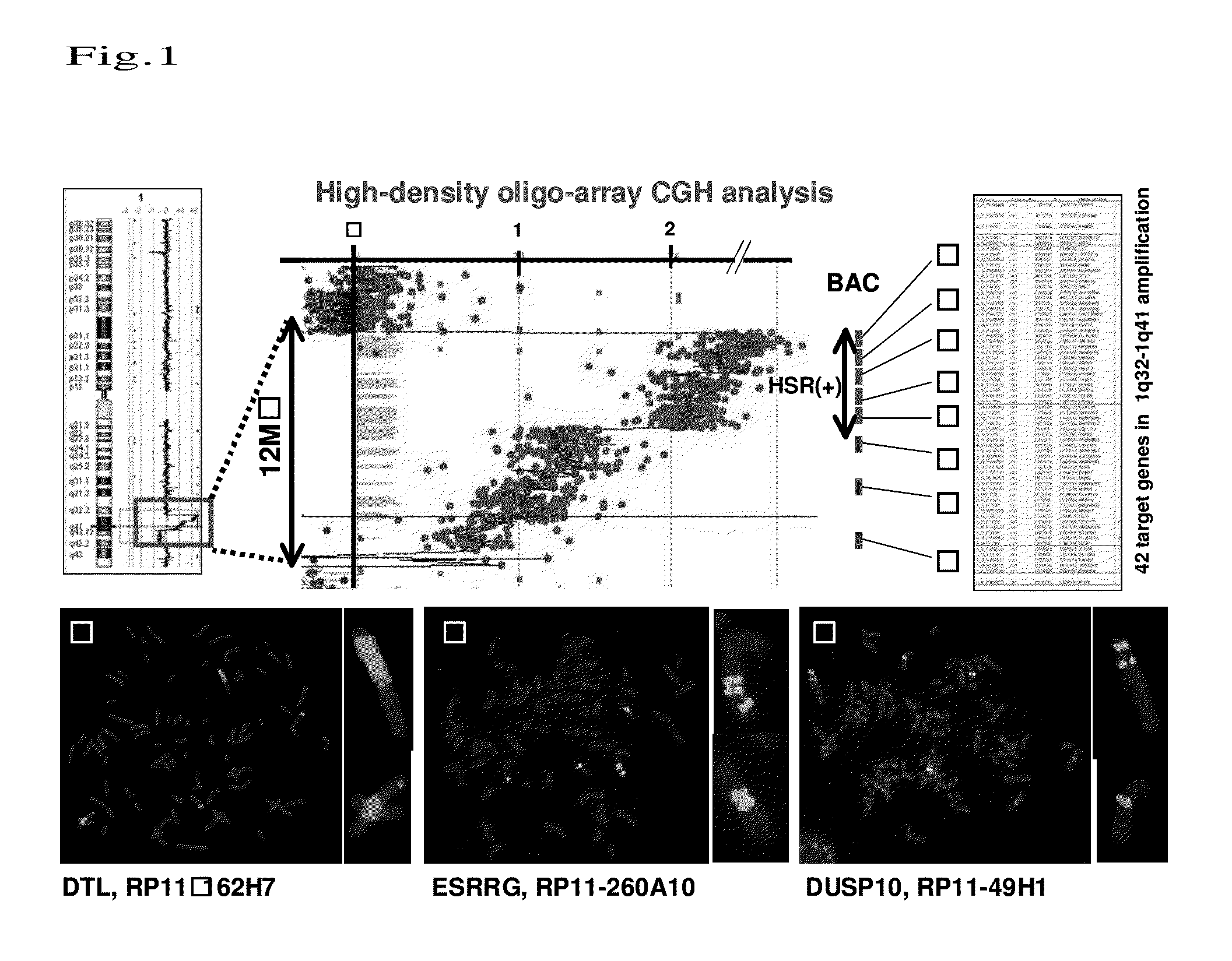
[0089]For detection of new genetic changes in esophageal carcinoma, an approximately 12-MB amplification region located at 1q32-1q41 (among known amplification regions of ESCC cell lines disclosed in the CGH data base Japan (http: / / www.cghtmd.jp / CGHDatabase / )) was mapped by high-density oligo array (Agilent 244 K high-density oligo-array) CGH analysis and the FISH method using genomic DNAs prepared from the above 43 types of ESCC cell line. Thus, 42 candidate genes within the region were identified (FIG. 1A). Specifically, in the region with the highest number of copies among slight changes in the number of copies as evaluated using the oligo array, an HSR (Homogeneously Staining Region) pattern was detected in the regions ranging from circled number 1 to circled number 4 even by the FISH method. In BAC regions with circled number 6 and circled number 8, changes in signal pattern in correlation with decreases in the nu...
example 2
Quantitative Analysis Regarding mRNA Expression of the Above 22 Genes
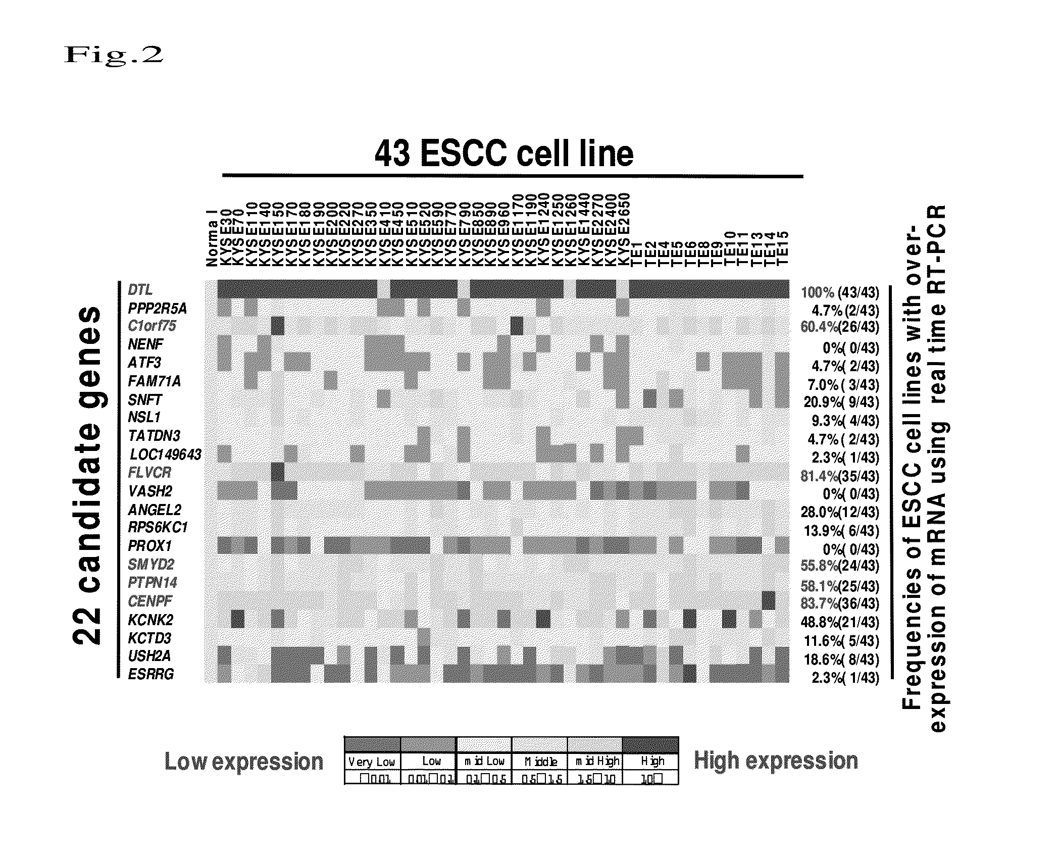
[0099]With use of the above 43 ESCC cell lines, quantitative analysis was conducted regarding mRNA expression of 22 types of gene selected in Example 1. An epithelium of a normal esophagus was used to represent a control expression level in quantitative RT-PCR. Total RNA was collected from each cell at the logarithmic growth phase and then cDNA was constructed by a standard method. cDNA was subjected to measurement of mRNA expression level by a quantitative real-time fluorescence detection method (ABI PRISM 7500 sequence detection System; Applied Biosystems, Foster City, Calif., U.S.A.) using protocols of TaqMan Gene Expression Assays (ABI, Applied Biosystems) and primers specific to each gene.
[0100]FIG. 2 shows the results. In FIG. 2, gene names are shown on the left, cell line names are shown on the top, and frequencies of cell lines exhibiting high-level expression in the normal tissues are shown on the right. B...
example 3
Knockdown Experiment Using siRNA of the Above 10 Genes
[0101]Knockdown was carried out using various siRNAs of the 10 types of gene in Example 2 and then cell growth assay was carried out. Specifically, amplification cell lines were subjected to analysis using an siRNA of Santa Cruzs (Santa Cruz Biotechnology, Inc.), Dharmacon (Lafayette, Colo., USA), or Sigma (Tokyo, Japan). Transfection was carried out using Lipofectamine 2000 (Invitrogen, St. Louis, Mo., U.S.A.) in reference to the protocols attached to siRNA (Santa Cruzs 10 nmol / L, Dharmacon 20 nmol / L, or Sigma 50 nmol / L). The degree of suppression of growth was evaluated using WST assay (colorimetric water-soluble tetrazolium salt assay).
[0102]FIG. 3 shows the results. The mRNA expression levels were shown on the right in FIG. 3. Knockdown by siRNAs was confirmed by quantitative analysis conducted for expression. Particularly C1orf75 and SMYD2 were found to exert significant effects of suppressing growth at 72 hours (FIG. 3).
PUM
| Property | Measurement | Unit |
|---|---|---|
| concentration | aaaaa | aaaaa |
| concentration | aaaaa | aaaaa |
| diameter | aaaaa | aaaaa |
Abstract
Description
Claims
Application Information
 - R&D
- Intellectual Property
- Life Sciences
- Materials
- Tech Scout
- Unparalleled Data Quality
- Higher Quality Content
- 60% Fewer Hallucinations
Browse by: Latest US Patents, China's latest patents, Technical Efficacy Thesaurus, Application Domain, Technology Topic, Popular Technical Reports.
© 2025 PatSnap. All rights reserved.Legal|Privacy policy|Modern Slavery Act Transparency Statement|Sitemap|About US| Contact US: help@patsnap.com


