Methods and kits for detecting misfolded proteins

a technology for misfolded proteins and kits, applied in the field of methods and kits for detecting misfolded proteins, can solve the problems of insufficient insight into the likelihood of the most severe symptoms developing, the method is not precise, and the risk of preeclampsia is low, so as to reduce the rate of abnormal folding and the effect of aggregation

Active Publication Date: 2016-01-05
RGT UNIV OF CALIFORNIA +1
View PDF8 Cites 16 Cited by
  • Summary
  • Abstract
  • Description
  • Claims
  • Application Information

AI Technical Summary

Benefits of technology

[0018]In a specific embodiment, the method of diagnosing or aiding in diagnosing preeclampsia in a pregnant woman comprises: (a) obtaining a urine sample or placental tissue sample from the pregnant woman; (b) combining the sample obtained in (a) with a dye that stains at least one protein or protein fragment present in the abnormal protein aggregates (e.g., Congo Red), under conditions under which the dye stains protein in the sample, thereby producing a urine sample or a placental tissue sample that further comprises the dye; and (c) analyzing the urine sample or placental tissue sample produced in (b) for the presence of (determining if the sample contains) supramolecular aggregates, (abnormal protein aggregates) associated with preeclampsia stained with the dye, wherein if the sample contains supramolecular aggregates associated with preeclampsia stained with the dye, it is indicative of preeclampsia in the woman and the woman is diagnosed as having or likely to have preeclampsia. If the sample contains such stained misfolded protein aggregates, she is more likely to have preeclampsia than if the sample does not contain abnormal protein aggregates associated with preeclampsia stained with the dye. The presence of abnormal protein aggregates associated with preeclampsia stained with the dye indicates that the woman has preeclampsia or is likely to have preeclampsia. Her status (having preeclampsia or not having preeclampsia (can be further assessed, using known methods, such as methods described herein, in order to confirm the determination resulting from the present method. The abnormal protein aggregate detected can be the abnormal protein aggregate described herein, which comprises at least one (one or more) of SerpinA1, ceruloplasmin, heavy-chain IgG, light-chain IgG, interferon inducible protein 6-16 or a fragment of any of the foregoing. Detection of stained abnormal protein aggregates can be carried out using known methods, such as dot blot analysis or simple visualization of an area of a stained surface (e.g., on a surface, such as filter paper) that comprises the urine or placental tissue sample combined with the dye (e.g., urine combined with Congo Red).
[0098]53. The inhibitor of paragraph 50, which stabilizes the native conformation of the protein, thereby decreasing the rate of abnormal folding and consequent aggregation.

Problems solved by technology

Currently, there is no single test to predict or diagnose preeclampsia or to foretell the severity of the condition that will develop in a particular patient.
These methods are imprecise and provide little insight into the likelihood of the most severe symptoms developing.
Moreover, the current diagnostics require physician oversight and invasive methodologies, further delaying and complicating early and immediate assessment.

Method used


Image

  • Methods and kits for detecting misfolded proteins
  • Methods and kits for detecting misfolded proteins
  • Methods and kits for detecting misfolded proteins

Examples


example 1

Methods

Protocol Congo Red Dot Blotting of Urine Samples (Red Dot Test):

[0244]1. Centrifuge obtained urine samples at 1500×g, 15 minutes and 4° C.[0245]2. Measure urine protein in the spun urine using the Pierce BCA kit (Thermo Scientific Cat# 23225). Urine samples may require a 12-fold dilution for protein measurement. Urine samples from women that are potentially preeclamptic have wide variations in protein concentration. Protein levels in urine also vary widely in healthy people depending on the state of hydration. For this reason it is preferred to equalize the total protein concentration. Out of over 680 urine samples obtained from women only approximately 5% (37 / 681) of urine samples measured [0246]3. Add the volume of sample that contains 200 μg protein to a conical centrifuge tube and concentrate to dryness using, e.g., a SPEEDVAC®.[0247]4. Prepare the Congo Red stock solution (5 mg / ml-) Congo red (Sigma, Cat#C6277) in water. Centrifuge at 14,000×g, 10 minutes to spin down un...

example 2

Methods

Study Design:

[0273]347 pregnant women were enrolled prospectively in the following groups: normotensive controls (CRL n=98, GA:27[7-42 weeks]); chronic hypertension (cHTN n=40, GA:32[11-41 weeks]), gestational hypertension (gHTN n=8 GA:37[26-39 weeks]), mild PE (mPE n=36, GA:36 [24-41 weeks]), severe PE (sPE n=117, GA:32[22-42 weeks]), superimposed PE (spPE n=33 GA:33[18-40 weeks]). 35 asymptomatic women were tested serially throughout gestation. A “Congo Red (CR) Dot” test was standardized with equal urine protein and objectively quantified within minutes as % Congo Red retention (CRR). CRR was evaluated for its ability to predict an indicated delivery for PE (IND) compared to protein-to-creatinine ratio (P / C) and the previously validated urine sFlt1 / PlGF ratio, as described herein and also in (U.S. Publ. No: US-2006-0183175; PCT / US2005 / 047010).

Results:

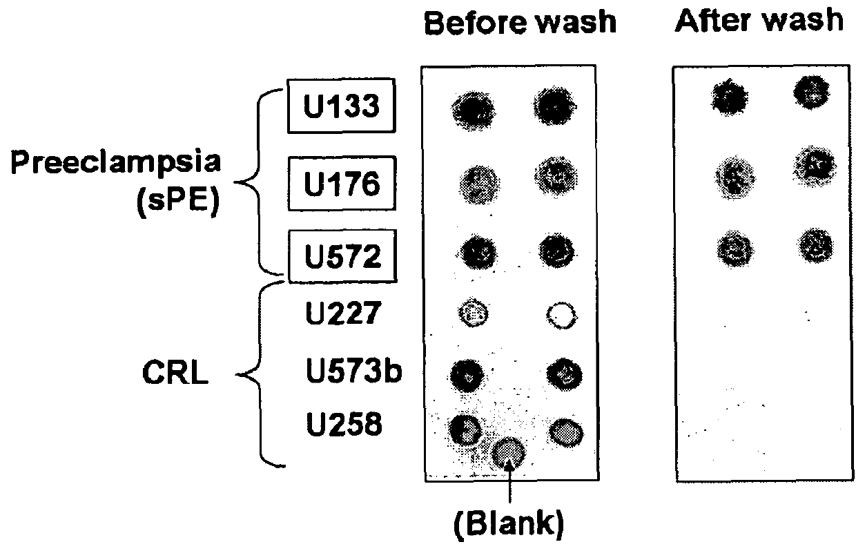
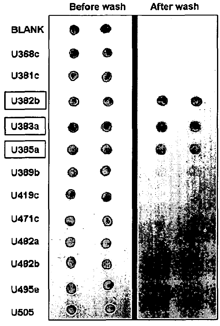
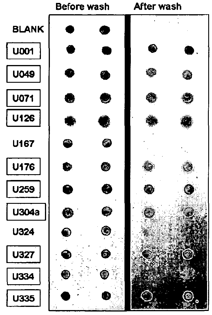
[0274]PE is characterized by increased excretion of misfolded proteins with affinity for the azo dye Congo Red (CR), also us...

example 3

Methods

Study Design:

[0276]111 urine samples from women enrolled in 3 groups: sPE (n=49, GA: 28±1 weeks), chronic hypertension (cHTN n=12, GA: 29±1 weeks) and normotensive controls (CRL n=50, GA: 28±1 weeks) were analyzed. Equal amounts of urine protein was subjected to dot blot using 3 conformation-specific antibodies recognizing prefibrillar soluble oligomers (A11, Invitrogen), ring-shaped protofibrils (Officer) or fibrils (OC). Specificity was confirmed by omitting the primary antibodies. Identity of aggregated component proteins was sought by mass spectrometry and validated by Western blot with sequence-specific antibodies.

Results

[0277]Protein conformational disorders such as Alzheimer's, light chain amyloidosis and prion diseases are propagated by amyloid fibril formation and aggregation due to defective folding of cellular proteins into aberrant 3D structures. Soluble pre-amyloid oligomers (intermediates in fibril assembly) have proteotoxic effects leading to endothelial damage...


PUM

PropertyMeasurementUnit
ODaaaaaaaaaa
pHaaaaaaaaaa
temperatureaaaaaaaaaa

Abstract

Methods, kits and compounds are provided that relate to the diagnosis, treatment, and / or prevention of preeclampsia.

Description

RELATED APPLICATIONS[0001]This application is a national stage filing under 35 U.S.C. §317 of international Application PCT / US2009 / 005957, filed Nov. 2, 2009, which designated the United States of America, and was published in English. This application claims the benefit of U.S. Provisional Application No. 61 / 197,914, filed on Oct. 31, 2008 and entitled “Preeclampsia is a disease characterized by specific supramolecular aggregates of misfolded protein” and U.S. Provisional Application 61 / 206,534, filed Jan. 29, 2009 and entitled “Preeclampsia is a disease characterized by specific supramolecular aggregates of misfolded proteins detected by antibodies that recognize specific conformations of protein oligomers.” The entire teachings of the referenced applications are incorporated herein by reference.BACKGROUND OF THE INVENTION[0002]Preeclampsia complicates 6-8% of pregnancies (Hauth, J. C. et al., Obstet Gynecol. 95(1):24-8, 2000) with an incidence in the US of 23.6 cases per 1000 del...

Claims


Application Information

Patent Timeline
Patent Type & Authority Patents(United States)
IPC IPC(8): G01N33/52G01N33/68
CPCG01N33/689G01N33/52G01N33/6803G01N2570/00G01N2800/50G01N2800/368G01N2333/90287G01N2333/8125G01N33/6866G01N33/6857G01N33/53G01N33/68G01N2333/4703
Inventor BUHIMSCHI, CATALIN S.BUHIMSCHI, IRINAGLABE, CHARLES G.
Owner RGT UNIV OF CALIFORNIA
Why Eureka
Try Eureka