
Tenocyte containing bioscaffolds and treatment using the same

a technology of tenocytes and bioscaffolds, applied in the field of preparing bioscaffolds, can solve the problems of low success rate of revision surgery for failed rotator cuff repair, significant morbidity associated with large and chronic tears, and the failure of rotator cuff repair devices to show long-term success,

Active Publication Date: 2016-10-11
ORTHOCELL PTY LTD
View PDF30 Cites 1 Cited by
  • Summary
  • Abstract
  • Description
  • Claims
  • Application Information

AI Technical Summary

Benefits of technology

The present invention provides a novel cell-based tissue engineering approach for the treatment of rotator cuff tear in mammals. The method involves the selective expansion of tenocytes in vitro, the seeding of a bioscaffold with the expanded tenocytes, and the implantation of the bioscaffold into the rotator cuff tear. The use of this approach has been shown to promote tendon healing and restore function in a rodent model of rotator cuff tear. The invention also provides a bioscaffold comprising cells, wherein the cells are pre-dominantly tenocytes. The bioscaffold may be made of a matrix, membrane, microbead, fleece, thread, or gel, and / or mixtures thereof.

Problems solved by technology

While surgical repair usually achieves high levels of functional improvement and patient satisfaction in the early post-operative stage, there is significant morbidity associated with large and chronic tears (Hattrup, (1995) J.
In addition, the success rate of revision surgery for failed rotator cuff repair is very low (Bigliani at al.
However, devices for the treatment of rotator cuff tear have generally failed to show successful long-term results with failure commonly occurring due to the invasion of non-functional fatty tissue, tenosynovitis, loosening or implant failure.
Further, continuous loading of the device and abrasion against joint tissues causes wear, creep and fatigue of the device and ultimately the device fails.
As such, the augmentation of surgical repair with devices such as bioscaffolds in rotator cuff tears has not been shown to improve the clinical outcome of patients (Iannotti, (1994) J. Am. Acad. Orthop. Surg. 2(2):87-95).

Method used


Image

  • Tenocyte containing bioscaffolds and treatment using the same
  • Tenocyte containing bioscaffolds and treatment using the same
  • Tenocyte containing bioscaffolds and treatment using the same

Examples


example 1

Animals and Bioscaffolds

[0097]Fifty Albino New Zealand White (Oryctolagus cuniculus) rabbits between 12 to 20 weeks old with body weight between 3-5 kg were used in this study. All rabbits were bred from an out bred rabbit colony maintained in the animal house facility of the University of Western Australia (Nedlands, Australia). Rabbits were fed ad libitum with rabbit / guinea pig pellets, hay, and provided water ad libitum. Rabbits were held in cages measuring 1.5 m wide, 0.75 m long, and 0.75 m high, with grid floors to prevent pod dermatitis. All operative procedures and cage activities were conducted under strict guidelines detailed by the National Health and Medical Research Council (NHMRC, Can berra, Australia).

[0098]Porcine-derived type I / III collagen bioscaffold (ACI Maix™) was supplied and manufactured by Matricel (Herzogenrath, Germany). The collagen bioscaffold is a white complex with two different sides: the rough side and the smooth side. The rough side appears as cross ...

example 2

Experimental Design

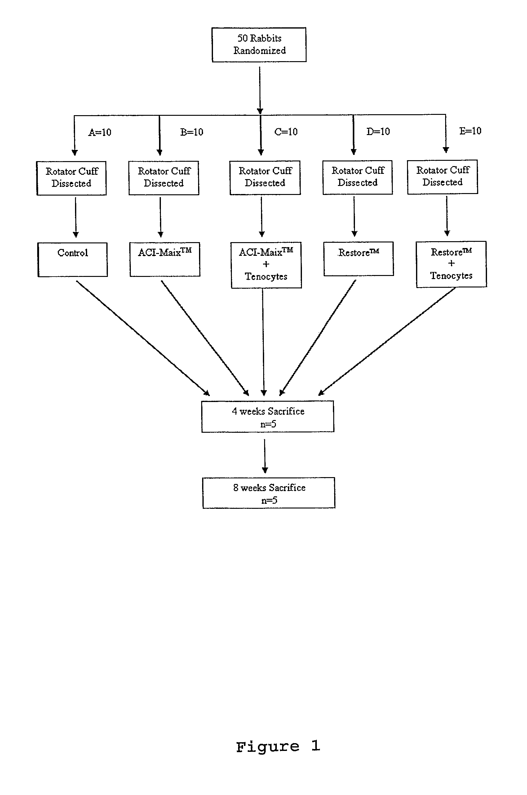
[0099]The fifty rabbits were randomly allocated into five groups of ten (FIG. 1). The left rotator cuffs were fully dissected and reconstructed by one of the five following methods: (1) Group A (Control treatment): The tendon excised during defect creation was in situ reimplanted into the defect immediately and sutured to the bone trough using 5-0 absorbable sutures; (2) Group B (ACI-Maix™): The defect was repaired by suturing ACI-Maix™ collagen bioscaffold as an interposition graft to the native tendon and bone trough borders; (3) Group C (ACI-Maix™ with autologous tenocytes): The cell-bioscaffold composite was used to repair the cuff defect in an identical manner to Group B; (4) Group D (Restore™): The cuff defect was repaired in an identical manner to Group B, but using Restore™ as an interposition graft; and (5) Group E (Restore™ with autologous tenocytes): The tenocytes seeded Restore™ composite was used to repair the cuff defect in an identical manner to Gro...

example 3

Harvest and Culture of Tenocytes

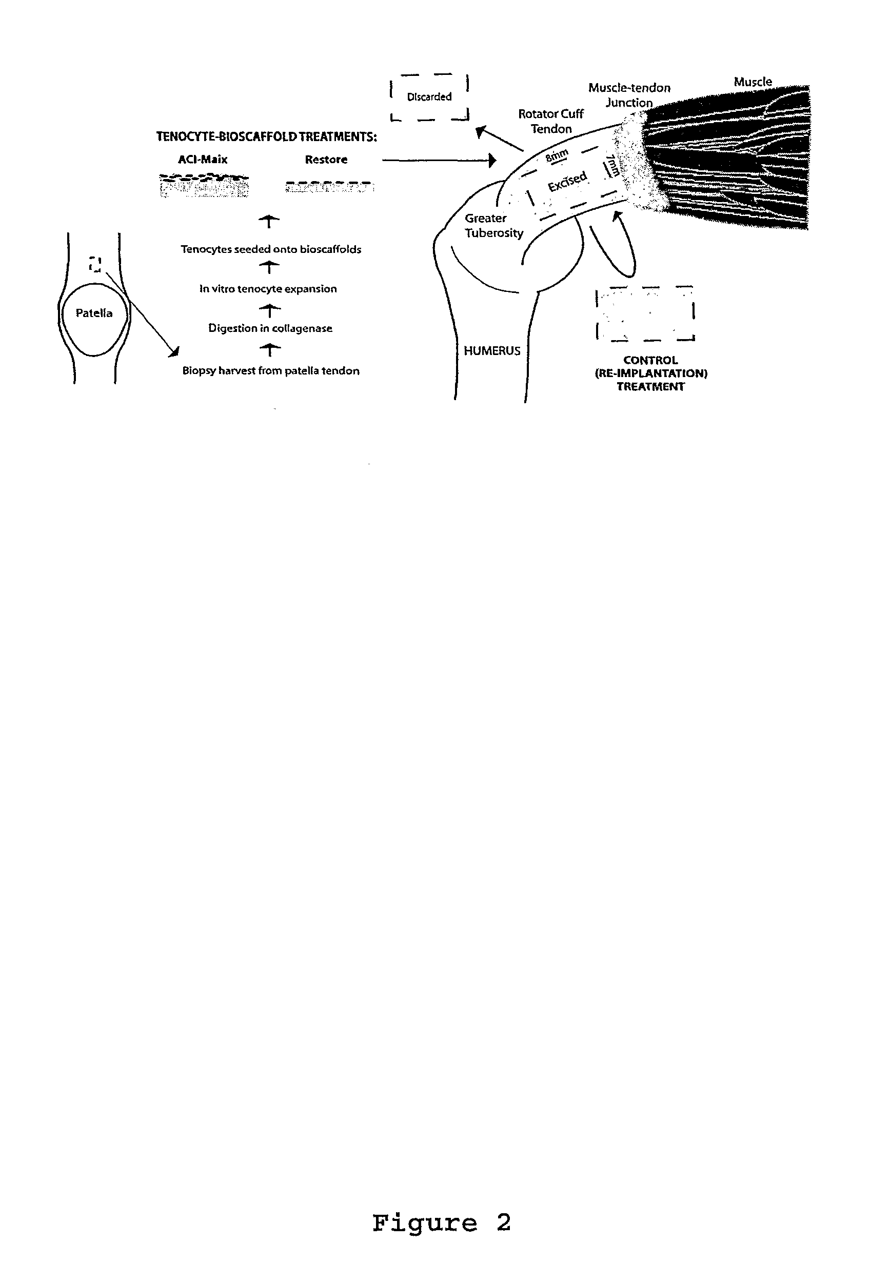
[0101]As shown in FIG. 2 tendon tissue of 3 mm diameter was obtained by biopsy punch from the rabbit patellar tendon and was washed in DMEM F-12 medium (GIBCO, Invitrogen, USA) supplemented with 10% fetal bovine serum (FBS), 100 μ / ml penicillin and 100 μg / ml streptomycin (hereafter medium refers to the same contents). The tendon tissue was then dissected into 0.5 mm of diameter and digested with collagenase (100 UI / ml Gibco, Invitrogen, USA) over night at 37° C. After the digestion, the solution was filter through a 0.22 μm filter to remove matrix debris and the cells released from tissue were centrifuged into pellet at 2000 rpm for 8 min. Supernatant containing enzymes were discarded and the pellet was resuspend in new medium. This washing process was repeated three times.

[0102]The cell pellet of resultant tenocytes was then resuspended in 5 ml of medium and placed into a culture flask at density between 103 to 104 cells / ml containing culture medium....


PUM

PropertyMeasurementUnit
body weightaaaaaaaaaa
pore sizeaaaaaaaaaa
thickaaaaaaaaaa

Abstract

The present invention relates to methods for preparing bio scaffolds useful in the repair of tears. More specifically, the invention relates to a method of treating rotator cuff tear in a mammalian subject in need thereof comprising the steps of: (i) selectively expanding tenocytes in vitro in culture medium comprising insulin or a functional derivative and a glucocorticoid or a glucocorticoid-like molecule to produce a culture of expanded tenocytes; (ii) seeding a bioscaffold with said expanded tenocytes to produce a tenocyte seeded bioscaffold; and (iii) implanting said tenocyte seeded bioscaffold proximal to the rotator cuff tear. The present invention also relates to a bioscaffold comprising cells, wherein more than 80% of said cells are tenocytes.

Description

CROSS REFERENCE TO RELATED APPLICATIONS[0001]This application is a U.S. national stage application of International Application No. PCT / AU2008 / 000583, filed Apr. 24, 2008, which claims the benefit of Australian Patent Application No. 2008901451, filed Mar. 26, 2008 and Australian Patent Application 2007902168, filed Apr. 24, 2007, all three of which are incorporated herein by reference in their entirety.FIELD[0002]The present invention relates to methods for preparing bioscaffolds useful in the repair of tears. More specifically, the present invention relates to the repair of rotator cuff tears using bioscaffolds seeded with tenocytes.INTRODUCTION[0003]Rotator cuff tendon tear is a common presentation resulting from injuries sustained from overhead activities (Briner et al., (1997) Sports Med. 24(1):65-71). Numerous studies have described different techniques for tendon repair. These include suture techniques (Gerber et al., (1999) J. Bone Joint Surg. Am. 81(9):1281-90), tendon to b...

Claims


Application Information

Patent Timeline
Patent Type & Authority Patents(United States)
IPC IPC(8): A61L27/38
CPCA61L27/386A61L27/3804A61L27/3895A61F2310/00365A61L2430/10A61P21/00A61L27/3662
Inventor ZHENG, MING-HAO
Owner ORTHOCELL PTY LTD
Learn More