System and method for imaging of vascular structures using non-contrast enhanced magnetic resonance imaging

a magnetic resonance imaging and enhanced technology, applied in the field of magnetic resonance imaging, can solve the problems of poor image quality, high cost of agents, and exposure of patients to added safety risks

Active Publication Date: 2016-11-29
NORTHSHORE UNIV HEALTHSYST
View PDF28 Cites 4 Cited by
  • Summary
  • Abstract
  • Description
  • Claims
  • Application Information

AI Technical Summary

Problems solved by technology

Such agents are costly and expose the patient to added safety risks, namely, nephrogenic systemic fibrosis.
However, at 1.5 Tesla Cartesian undersampling factors larger than two to four typically produce poor image quality.
Unfortunately, TOF produces nondiagnostic image quality outside of the head and neck.
However, the data must be sparse in order to minimize radial streak artifacts.
Furthermore, the most common way to acquire image sets suitable for subtraction is to acquire and subtract a contrast-enhanced and non-contrast-enhanced data set, which reintroduces the drawback of using contrast agents and the implication of nephrogenic systemic fibrosis.

Method used


Image

  • System and method for imaging of vascular structures using non-contrast enhanced magnetic resonance imaging
  • System and method for imaging of vascular structures using non-contrast enhanced magnetic resonance imaging
  • System and method for imaging of vascular structures using non-contrast enhanced magnetic resonance imaging

Examples


Embodiment Construction

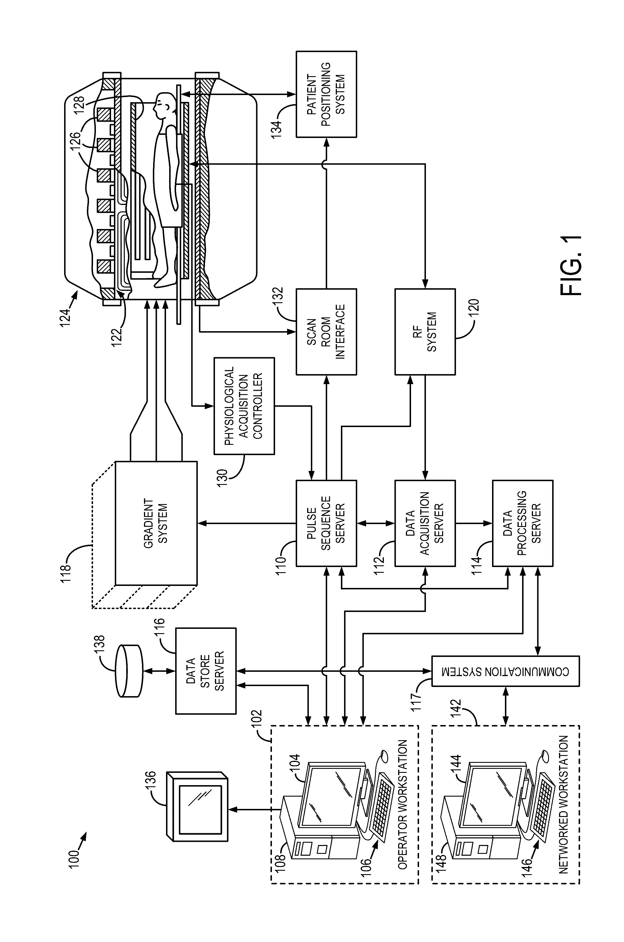
[0019]Referring particularly now to FIG. 1, an example of a magnetic resonance imaging (MRI) system 100 is illustrated. The MRI system 100 includes an operator workstation 102, which will typically include a display 104, one or more input devices 106, such as a keyboard and mouse, and a processor 108. The processor 108 may include a commercially available programmable machine running a commercially available operating system. The operator workstation 102 provides the operator interface that enables scan prescriptions to be entered into the MRI system 100. In general, the operator workstation 102 may be coupled to four servers: a pulse sequence server 110; a data acquisition server 112; a data processing server 114; and a data store server 116. The operator workstation 102 and each server 110, 112, 114, and 116 are connected to communicate with each other. For example, the servers 110, 112, 114, and 116 may be connected via a communication system 117, which may include any suitable n...


PUM


Abstract

A system and method is provided for acquiring a medical image of a portion of a vascular structure of a subject using a magnetic resonance imaging (MRI) system. At least one radio frequency (RF) saturation pulse is applied to a selected region of a subject that is free of exogenous contrast agents using the MRI system to saturate spins of all tissues within the selected region. A delay time is selected to allow an inflow of unsaturated vascular spins that are free of influence from exogenous contrast agent into the selected region through vascular structures within the selected region. A sparse dataset is formed from a series of spatially-encoded views from the selected region using an undersampled radial k-space trajectory in which the center of k-space is sampled for each view in the series of spatially-encoded views. An image of the vascular structures within the selected region is reconstructed from the sparse dataset.

Description

CROSS-REFERENCE TO RELATED APPLICATIONS[0001]The present application is based on, claims priority to, and incorporates herein by reference in its entirety U.S. Provisional Application Ser. No. 61 / 703,040, filed Sep. 19, 2012, and entitled, “SYSTEM AND METHOD FOR IMAGING OF VASCULAR STRUCTURES USING NON-CONTRAST ENHANCED MAGNETIC RESONANCE IMAGING.”BACKGROUND OF THE INVENTION[0002]The invention relates to a system and method for performing magnetic resonance imaging and, more particularly, to a system and method for acquiring images using magnetic resonance imaging that provide improved, clinically-valuable, magnetic-resonance images of vascular structures.[0003]When a substance such as human tissue is subjected to a uniform magnetic field (polarizing field B0), the individual magnetic moments of the nuclear spins in the tissue attempt to align with this polarizing field, but precess about it in random order at their characteristic Larmor frequency. Usually the nuclear spins are comp...

Claims


Application Information

Patent Timeline
Patent Type & Authority Patents(United States)
IPC IPC(8): G01R33/563G01R33/48G01R33/483
CPCG01R33/5635G01R33/4824G01R33/4838
Inventor EDELMAN, ROBERT R.KOKTZOGLOU, IOANNIS
Owner NORTHSHORE UNIV HEALTHSYST
Why Eureka
Try Eureka