Eureka AIR delivers breakthrough ideas for toughest innovation challenges, trusted by R&D personnel around the world.

Implantation of a cardiac xenograft from a B4GALNT2KO and GTKO transgenic pig to reduce immunogenicity

a transgenic pig and xenograft technology, which is applied in the field of implantation of cardiac xenografts from b4galnt2ko and gtko transgenic pigs to reduce immunogenicity, can solve the problems of devastating hyperacute rejection of grafts usually within hours, and the shortage of organs for transplantation, so as to reduce the immunogenicity of pigs, prolong the durability of xenografts

Active Publication Date: 2017-05-09
MAYO FOUND FOR MEDICAL EDUCATION & RES
View PDF24 Cites 1 Cited by
  • Summary
  • Abstract
  • Description
  • Claims
  • Application Information

AI Technical Summary

Benefits of technology

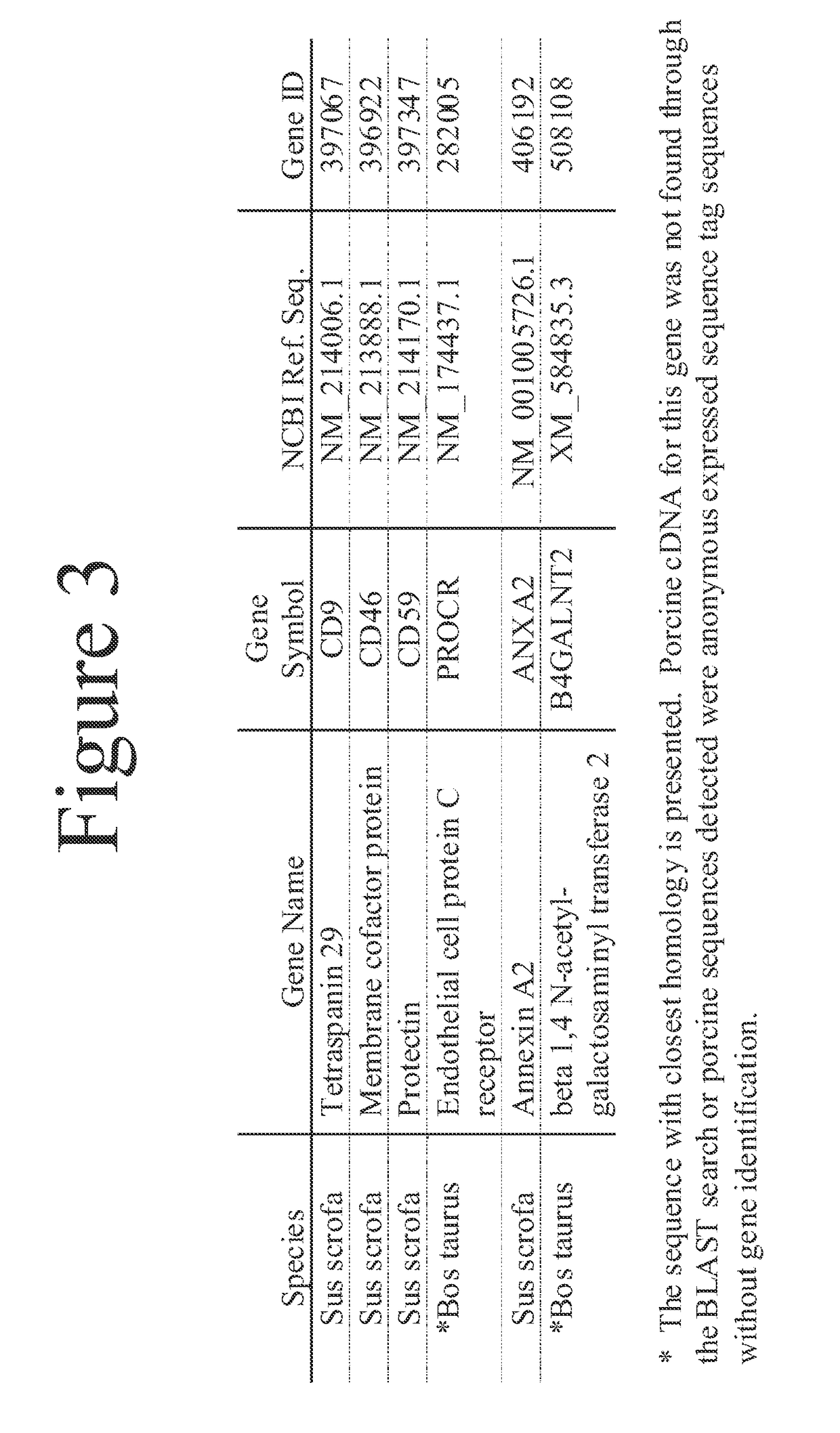
[0007]This document provides methods and materials for reducing cardiac xenograft rejection. For example, this document provides methods and materials for preparing transgenic pigs expressing reduced or no endogenous Sda or SDa-like glycans produced from a porcine β1,4 N-acetyl-galactosaminyl transferase 2 (B4GALNT2) glycosyltransferase and reduced or no endogenous α-Gal antigens produced from the porcine α1-3 galactosyl transferase (GT) glycosyltransferase, methods and materials for modifying a xenograft recipient's immunological response to non-Gal antigens (e.g., CD46, CD59, CD9, PROCR and ANXA2) to reduce cardiac xenograft rejection, and methods and materials for monitoring the progression of, if any, xenotransplant immunologic rejection. In some cases, this document provides methods for implanting a pig xenograft heart into a human. The pig xenograft donor can be a pig that contains genetic disruptions in α1-3 galactosyl transferase (GT) nucleic acid and β1,4 N-acetyl-galactosaminyl transferase 2 (B4GALNT2) nucleic acid. Such pigs can lack the ability to express Sda or SDa-like glycans and α-Gal antigens. The methods and materials described herein can be used to reduce immunogenicity of the pig to primate cardiac xenograft upon implantation and prolong the durability of the xenograft. This can benefit patients in chronic heart failure on the heart transplant waiting list for a donor heart.

Problems solved by technology

There is a chronic shortage of organs for transplantation.
Xenotransplantation can be limited by an immunological rejection of the transplanted organ.
The combination of abundant α-Gal antigen in pig organs and high levels of preformed anti-Gal antibody in nonhuman primates (a model for humans) can result in a devastating hyperacute rejection of the graft usually within hours.
Such pigs can lack the ability to express Sda or SDa-like glycans and α-Gal antigens.

Method used


Image

  • Implantation of a cardiac xenograft from a B4GALNT2KO and GTKO transgenic pig to reduce immunogenicity
  • Implantation of a cardiac xenograft from a B4GALNT2KO and GTKO transgenic pig to reduce immunogenicity
  • Implantation of a cardiac xenograft from a B4GALNT2KO and GTKO transgenic pig to reduce immunogenicity

Examples


example 1

Identifying Non-Gal Antigens Present on the Endothelial Cell Membranes of Pig Hearts Involved in Immunologic Xenograft Rejection

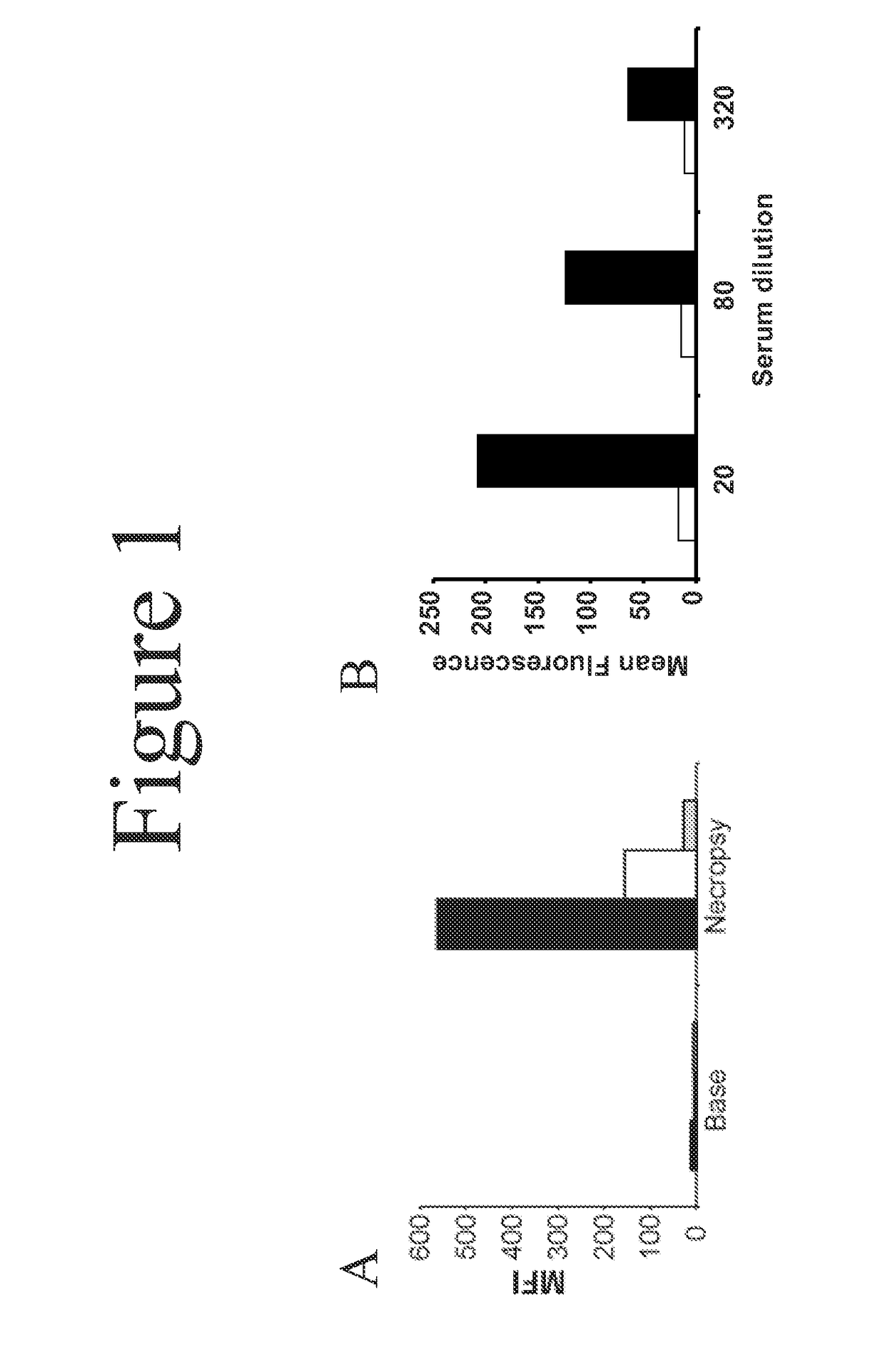
[0045]Heterotopic pig-to-primate cardiac xenografts were performed using GT-positive and GTKO donor hearts without immunosuppression. See Davila et al., Xenotransplantation, 13(1):31-40 (2006). Sera obtained at necropsy was screened by flow cytometry to measure IgG binding to GTKO porcine aortic endothelial cells (PAECs). Serum from both GT-positive and GTKO recipients showed an induced antibody response to non-Gal antigens as evidenced by increased IgG binding to GTKO PAECs in necropsy sera compared to pre-transplant sera (FIG. 1). This necropsy sera was used for the library screen.

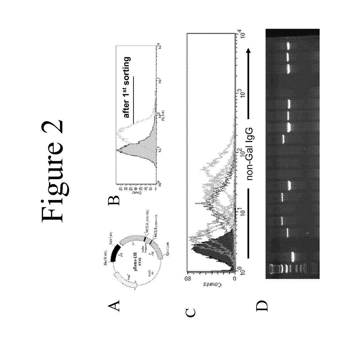
[0046]A standard cDNA expression library was produced using mRNA from GT-positive and GTKO porcine aortic endothelial cells. This library (pRETRO-PAEC) was made in the pRetro-LIB vector (Clontech, FIG. 2A) and was co-transfected with pVSV-G (encoding an envelope glycoprotein fro...

example 2

Engineering a Targeted Disruption in the Porcine B4GALNT2 Nucleotide Sequence

[0052]The cDNA sequence of porcine B4GALNT2 and its conservation in human and mouse provides the needed information to design a targeting vector suitable for disrupting the porcine B4GALNT2 gene using the standard methods of homologous recombination. The amino acids encoded by the nucleotides of individual exons in the human and murine genes show conservation (FIG. 6). This is a common observation in mammalian genes when exons from different species encode the same or similar portions of a polypeptide. This conservation suggests that the porcine B4GALNT2 gene is made up of 11 coding exons and suggests the approximate location for exon boundaries within the porcine cDNA.

[0053]The porcine, human and murine B4GALNT2 cDNA sequences exhibit a high level of conservation in the region that encodes the C-terminal portion of the polypeptide. This region of the human B4GALNT2 cDNA sequence encodes a portion of the B4...

example 3

Monitoring the Progress of Pig Heart Xenograft Rejection

[0056]Utilizing cDNAs encoding the polypeptide non-Gal antigens identified herein, HEK cell lines were developed expressing each of the polypeptide non-Gal antigens with the exception of B4GALNT2. These cell lines were directly used to monitor non-Gal antibody responses to individual non-Gal antigens (i.e. CD46, CD59, CD9, PROCR and ANXA2). Stable HEK cell lines expressing each of these cDNAs were incubated with pre-transplant and necropsy serum (diluted 1:40) from a non-immunosuppressed heterotopic cardiac xenograft recipient. Antibody binding was detected using a FITC conjugated anti-human IgG. Flow cytometry was used to determine the antibody response to each of the antigens (FIG. 12). The xenograft recipient had a strongly induced antibody response to the B4GALNT2 antigen and PROCR, a less intense though positive response to porcine CD9 and CD46 and a minimal response to porcine CD59 and ANXA2. This same strategy can be use...


PUM

PropertyMeasurementUnit
durabilityaaaaaaaaaa
acidicaaaaaaaaaa
genomic structureaaaaaaaaaa

Abstract

This document provides methods and materials involved in reducing cardiac xenograft rejection. For example, methods and materials for preparing transgenic pigs expressing reduced or no endogenous Sda or SDa-like glycans derived from the porcine β1,4 N-acetyl-galactosaminyl transferase 2 (B4GALNT2) glycosyltransferase and / or reduced or no endogenous α-Gal antigens, methods and materials for modifying the xenograft recipient's immunological response to non-Gal antigens (e.g. CD46, CD59, CD9, PROCR, and ANXA2) to reduce cardiac xenograft rejection, and methods and materials for monitoring the progress of xenotransplant immunologic rejection are provided.

Description

CROSS-REFERENCE TO RELATED APPLICATIONS[0001]This application is a National Stage application under 35 U.S.C. 371 of International Application No. PCT / US2011 / 031976, having an International Filing Date of Apr. 11, 2011, which claims the benefit of priority to U.S. Provisional Application Ser. No. 61 / 332,127, filed May 6, 2010. The disclosure of the prior application is considered part of (and is incorporated by reference in) the disclosure of this application.STATEMENT AS TO FEDERALLY SPONSORED RESEARCH[0002]This invention was made with government support under AI066310 awarded by the National Institutes of Health. The government has certain rights in the invention.BACKGROUND[0003]1. Technical Field[0004]This document relates to methods and materials involved in reducing cardiac xenograft rejection. For example, this document provides methods and materials for preparing transgenic pigs expressing reduced or no endogenous Sda, reduced or no endogenous SDa-like glycans derived from th...

Claims


Application Information

Patent Timeline
Patent Type & Authority Patents(United States)
IPC IPC(8): A01K67/027C12N15/00A61K35/34C12N15/85A61K39/00A61K38/00
CPCA61K39/001A01K67/0276A61K35/34C12N15/8509A01K2227/108A01K2267/02A01K2267/025A61K38/00A01K2207/30A61P37/06
Inventor MCGREGOR, CHRISTOPHER G. A.BYRNE, GUERARD W.
Owner MAYO FOUND FOR MEDICAL EDUCATION & RES
Why Eureka
Eureka Blog
Learn More