Device for reliably determining biometric measurement variables of the whole eye

a biometric and eye technology, applied in the field of eye biometric measurement variables, can solve the problems of low gain from scheimpflug anterior chamber measurement compared to additional costs, and the combination cannot measure all parameters of the eye, so as to achieve fast, movement-free measurement of the a-scan over the whole eye length

Active Publication Date: 2017-05-16
CARL ZEISS MEDITEC AG
View PDF23 Cites 1 Cited by
  • Summary
  • Abstract
  • Description
  • Claims
  • Application Information

AI Technical Summary

Benefits of technology

[0019]The multi-point keratometer ensures that, firstly, a sufficient number of keratometer points are available for measuring the corneal surface with a high resolution, but that the density of the measurement points is low enough for it to be possible to detect the Skrew rays. By contrast, the telecentricity ensures that the positioning inadequacies of the measurement instrument in relation to the eye to be measured do not lead to a local mismatch of the reflection points.
[0020]What the swept-source OCT scan, which captures the whole eye over the length thereof, achieves is that both anterior chamber and retina structures can be detected in the A-scan or B-scan and hence an orientation on the basis of the retina and the anterior chamber / cornea becomes possible during the scan. This makes it easier to combine the A- and / or B-scans to form a consistent whole-eye image. Here, the swept-source OCT is better than other OCT variants, such as time domain OCTs or spectrometer-based OCTs, in ensuring fast, movement artifact-free measurement of the A-scan over the whole eye length.

Problems solved by technology

Although Scheimpflug, PCI (partial coherence interference) and topography systems or combined instruments of same measure some of the aforementioned variables, these combinations cannot measure all parameters of the eye.
In particular, it is not possible to measure the lens rear side and the retina, or the respective profile of these areas, since these systems, even in a combined system, are restricted to measuring the anterior chamber and the axial one-dimensional length of the eye.
However, since the OCT can also detect the anterior chamber, the gain from the Scheimpflug anterior chamber measurement is low compared to the additional costs.
Compared to the topography / OCT combined systems, pure OCT systems are disadvantageous in that measuring the topography of the cornea by means of conventional topometers (in particular Placido systems) is substantially more accurate than the measurements of the OCT systems which are influenced by movement artifacts.
Although said OCT systems can reduce these movement artifacts by faster measurements or by measurements which are registered to the eye, this is only possible with significant outlay and not readily possible in a reliable enough manner.
However, the described device exhibits some significant disadvantages, which reduce the reliability of the measurement values.
Although Placido topography has a very high resolution, it is less reproducible in terms of reconstructing the surface when compared to keratometer measurements.
This is due, firstly, to the assumptions made during the reconstruction of the topography in order to achieve the high resolution and / or in the lacking telecentricity / insufficient focusability of many topography systems compared to keratometers, and so positioning errors of the measurement instrument in relation to the patient become relevant during the topography measurement.
Furthermore, Placido topographs do not allow so-called Skrew rays to be taken into account, which are always generated in the case of the Placido ring illumination when the cornea is curved not only in a central plane through the corneal vertex but also in a plane perpendicular thereto, i.e. if it has azimuthal curvature.
As a result of not taking this into account, the corneal surface is not reproduced correctly.
Thus, overall, a Placido topograph does not reproduce the radius or, in general, the front side of the cornea reliably enough as required for the IOL calculation.
Furthermore, time-domain OCT systems are too slow and competitively priced spectrometer-based systems do not have the axial resolution and / or have a too small axial scanning or detection depth such that the eye length does not occur with the resolution required for the IOL calculation or such that there are only partial depth measurements.
However, whole-eye biometrics, i.e. establishing the areas of the whole eye optically relevant to the visual faculty of the eye in terms of their position and their profile in the eye, are, in principle, possible in both cases by separate measurement of the anterior and posterior chamber and subsequent synthesis of the data, but the registration of the images to one another is often unreliable due to lack of a suitable common reference variable in the segment images.
Therefore, the Placido time-domain OCT system does not allow all biometric data to be obtained in a sufficiently reliable and simple manner for the IOL calculation.
However, the described device also exhibits some significant disadvantages, which reduce the reliability of the measurement values or leaves open important points which are relevant to whole-eye biometrics:
Furthermore, this does not solve the problem of assigning the topography measured by the keratometer to the spatial data from the OCT data, nor does it ensure that the OCT measurements are taken particularly quickly in order to compensate for the eye movement during the measurement.

Method used


Image

  • Device for reliably determining biometric measurement variables of the whole eye
  • Device for reliably determining biometric measurement variables of the whole eye
  • Device for reliably determining biometric measurement variables of the whole eye

Examples


Embodiment Construction

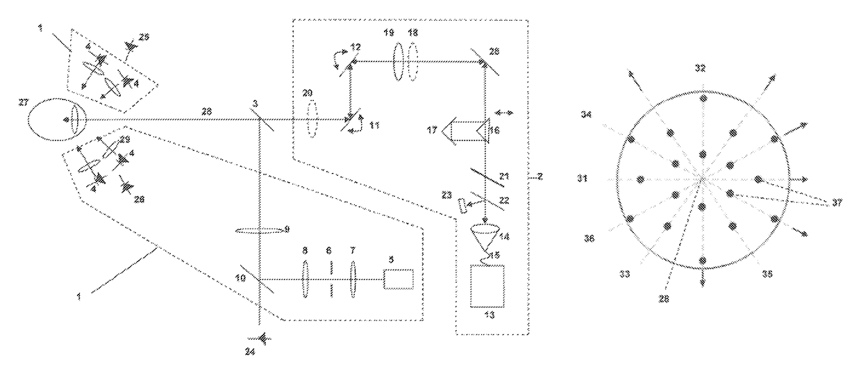
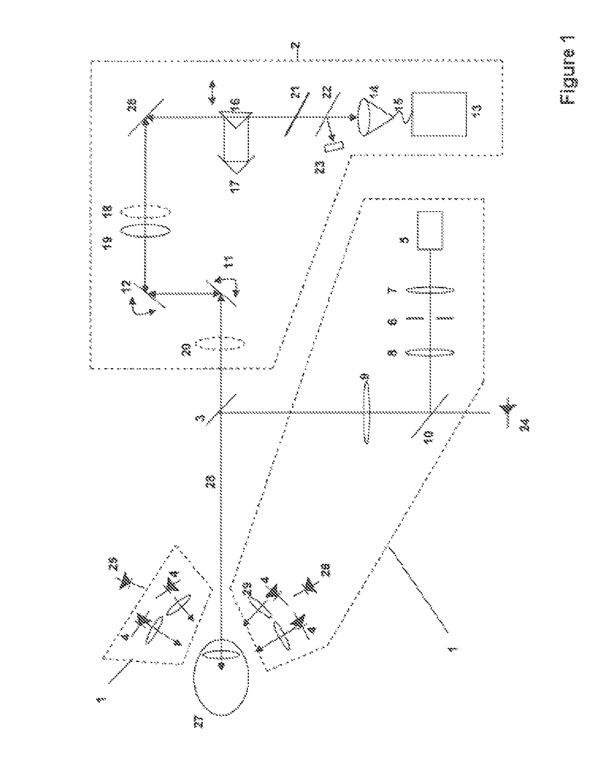
[0027]The device according to the invention for measuring biometric variables of the eye for calculating intraocular lenses consists of a multi-point keratometer and an OCT arrangement, wherein the multi-point keratometer is configured such that the keratometer measurement points are illuminated telecentrically and detected telecentrically and that the OCT arrangement is designed as a laterally scanning swept-source system with a detection region detecting the whole eye over the whole axial length thereof.

[0028]FIG. 1 shows a basic optical design of the device: by means of the beam splitter 3, an OCT system 2 is unified with a multi-point keratometer system 1 on a common instrument axis 28 such that both can biometrically measure the eye 27 in a laterally assignable manner.

[0029]Here, the multi-point keratometer system 1 consists of several light sources, preferably LEDs 4, at different radial distances from the instrument axis 28. Lens attachments 29 ensure that the LEDs telecentri...


PUM


Abstract

A device for determining biometric variables of the eye, as are incorporated in the calculation of intraocular lenses including a multi-point keratometer and an OCT arrangement. The keratometer measurement points are illuminated telecentrically and detected telecentrically and the OCT arrangement is designed as a laterally scanning swept-source system with a detection region detecting the whole eye over the whole axial length thereof. The multi-point keratometer ensures that a sufficient number of keratometer points are available for measuring the corneal surface. By contrast, telecentricity ensures that the positioning inadequacies of the measuring instrument in relation to the eye to be measured do not lead to a local mismatch of the reflection points. The swept-source OCT scan detects the whole eye over the length thereof so that both anterior chamber structures and retina structures can be detected and a consistent whole eye image can be realized.

Description

RELATED APPLICATIONS[0001]The present application is a National Phase entry of PCT Application No. PCT / EP2013 / 070199, filed Sep. 27, 2013, which claims priority from DE Patent Application No. 10 2012 019 474.9, filed Sep. 28, 2012, and also claims priority to U.S. Provisional Application No. 61 / 707,004 filed Sep. 28, 2012, said applications being hereby incorporated by reference herein in their entirety.FIELD OF THE INVENTION[0002]The present invention relates to a device for determining biometric variables of the eye, as are incorporated in the calculation of intraocular lenses. Such variables are the radii—including the orientation thereof—of the corneal front side and the corneal rear side, asphericity of the cornea, thickness of the cornea—in the center or else as one- or two-dimensional profile —, anterior chamber depth, lens thickness, radii of the lens front side and / or rear side, axis length of the eye, in general the position and shape of the optically effective interfaces ...

Claims


Application Information

Patent Timeline
Patent Type & Authority Patents(United States)
IPC IPC(8): A61B3/107A61B3/117A61B3/10
CPCA61B3/107A61B3/102A61B3/1005A61B3/1173A61B3/117
Inventor EBERSBACH, RALFHACKER, MARTINANTKOWIAK, GERARDKLOPFLEISCH, PETERBAJRAMOVIC, FERIDBUHREN, TOBIASREICH, MATTHIAS
Owner CARL ZEISS MEDITEC AG
Why Eureka
Try Eureka