
Method for detection, differentiation and quantification of T cell populations by way of reverse transcription quantitative real time PCR (RT-qPCR) technology

a t cell population and reverse transcription technology, applied in the field of methods for detection, differentiation and quantification of t cell populations, can solve the problems of inability to analyze the functionality of t cells, insufficient development of protective mechanisms, and routine monitoring of peptide mhc multimeres

Active Publication Date: 2017-08-15
LOPHIUS BIOSCI
View PDF25 Cites 0 Cited by
  • Summary
  • Abstract
  • Description
  • Claims
  • Application Information

AI Technical Summary

Benefits of technology

This approach enhances sensitivity and reliability in detecting activated T cells, enabling better discrimination between different T cell populations and aiding in the differential diagnosis of infectious and autoimmune diseases.

Problems solved by technology

Interference with the complex T cell function may lead to an erroneous activation of autoaggressive T cells and concomitantly elicit severe autoimmune diseases such as multiple sclerosis (MS), rheumatoid arthritis (RA) and juvenile diabetes (type I diabetes).
In autoimmune diseases these protective mechanisms are inhibited or are only insufficiently developed.
An analysis of the functionality of the T cells is not possible with this method.
The most important limitation in using peptide MHC multimeres for the routine monitoring of disease or pathogen specific T cells ensue because the MHC / peptide multimeres are epitope as well as HLA specific.
Thus, an exhaustive monitoring of pathogen as well as disease specific T cells in subjects with different HLA constellation requires the use of a broad spectrum of different peptide / MHC multimeres, which brings about high costs.
In addition, these peptide / MHC multimeres are so far only available for a limited spectrum of MHC molecules.
These available technologies allow the determination of disease and pathogen specific CD4+ or CD8+ memory T cells, but are not suitable or only very limited suitable for detection of activated T helper cells, which occur only transiently during the active course of disease.
However, for certain applications in which discrimination between activated and non-activated T cells is required, such as in the detection of activated T cells during acute microbial infections and reactivations or the detection of activated autoaggressive T cells in the case of suspected multiple sclerosis or type 1 diabetes, are methods which are based on in vitro restimulation of memory T cells not or only very limited employable.
The methods hitherto available for detection of activated CD4+ T cells exhibit so far only a low sensitivity and are thus disadvantageous.
However, this small number of available specific activated T cells hampers reliable and unambiguous detection which also satisfies diagnostic requirements, since the detection limit of these methods available hitherto is frequently undercut.
However, CD40 ligand is hardly detectable by flow cytometry, because the binding of antibodies leads to an internalisation of the CD40 ligand.
In contrast, the CD25 protein may not only be found on activated T-helper cells but also on regulatory T cells and is thus not suitable for a reliable distinction between these two subpopulations of T helper cells.
A further disadvantage of the methods hitherto known is, that the reliable detection of some marker proteins by way of ELISA, ELISpot or FACS technology is impossible due to the membrane localisation of the marker proteins, the presence of preformed marker proteins in intracellular vesicles, and the high unspecific reactivity of the available antibodies with cellular proteins.

Method used


Image

  • Method for detection, differentiation and quantification of T cell populations by way of reverse transcription quantitative real time PCR (RT-qPCR) technology
  • Method for detection, differentiation and quantification of T cell populations by way of reverse transcription quantitative real time PCR (RT-qPCR) technology
  • Method for detection, differentiation and quantification of T cell populations by way of reverse transcription quantitative real time PCR (RT-qPCR) technology

Examples


examples

[0143]In a preferred embodiment of the invention it is envisaged, that the kit comprises furthermore primer, which allow the detection of markers of the APC by PCR, qPCR or mircoarray.

[0144]In the following the invention is illustrated by the subsequent examples. These examples are to be considered as specific embodiments of the invention and shall not be considered to be limiting.

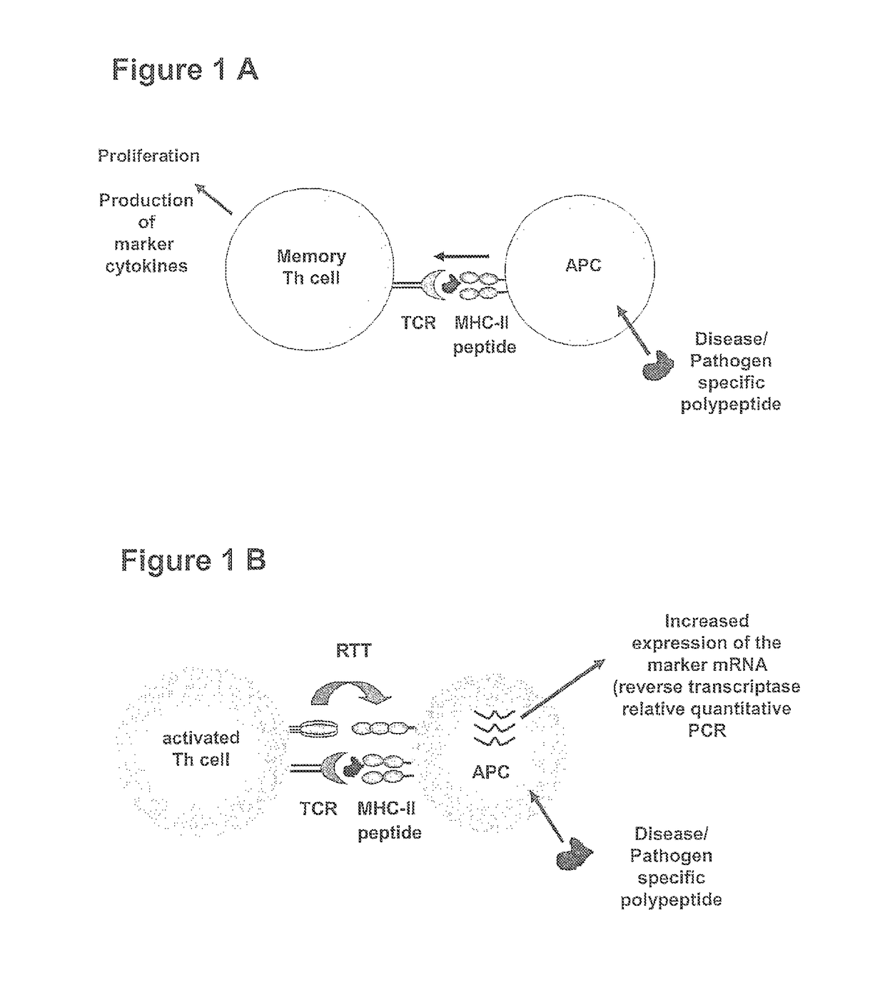
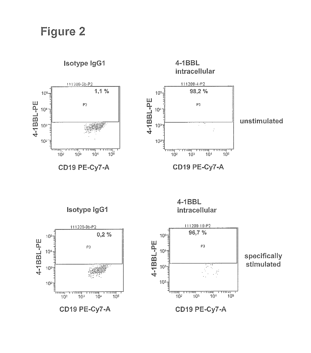
[0145]In order to establish the method for detection, differentiation and quantification of specific T cell populations, such as activated T cells, T cell induced maturation processes in antigen-presenting cells (APC) were analysed. For this purpose a cell culture model was developed, which is based on the coculture of antigen-loaded APC with ex vivo expanded, activated antigen-specific T helper cells. In this model system cocultures of (i) unloaded APCs or of APCs loaded with an irrelevant antigen with pre-activated antigen-specific T helper cells and (ii) antigen-loaded APCs with un-specifically activate...

example 2

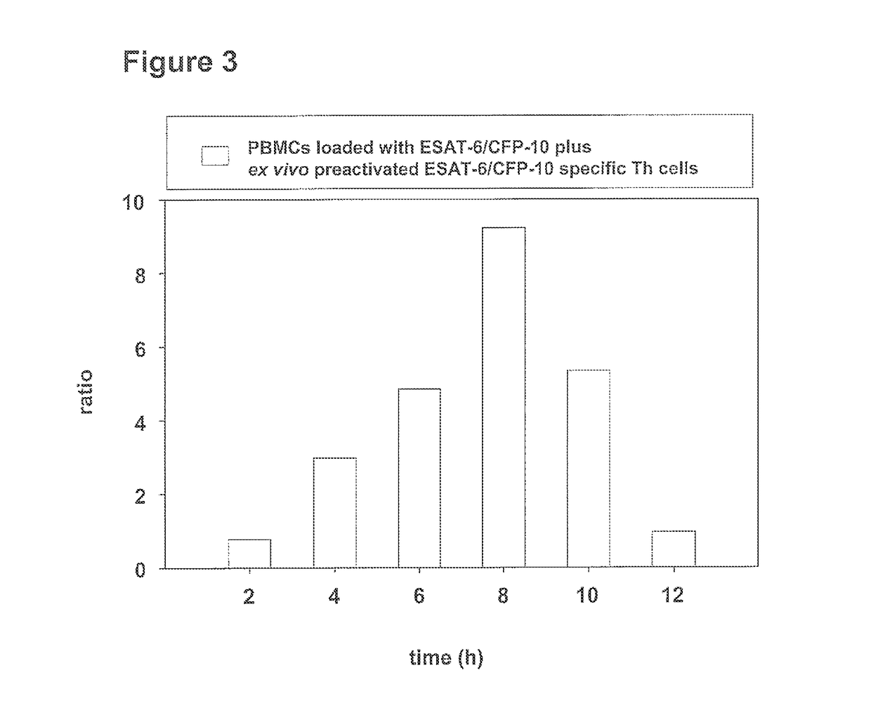
Correlation of Induction of 4-1BB Ligand mRNA Production in Cocultures of ESAT-6 / CFP-10 Protein-Loaded PBMCs with the Number of Ex Vivo Expanded ESAT-6 / CFP-10 Protein-Specific Th Cells

[0153]ESAT-6 / CFP-10 specific Th cells of subjects latently infected with Mycobacterium tuberculosis were expanded ex vivo as described in example 1 under item a) and were subsequently cocultured with freshly prepared autologous PBMCs as described in example 1 under item b). In this process the number of ESAT-6 / CFP-10 specific Th cells cocultured with 1×10⁶ PBMCs was titrated in a semi logarithmic concentration series (concentration steps: none, 117, 370, 1170, 3700, 11700, 37000, 117000 preactivated Th cells per 1×10⁶ PBMCs). After 0; 0.5; 1; 2; 3; 4; 6; 8 and 10 hours of incubation 2×10⁵ cells were removed, pelleted for 5 min at 300 g and the pellet was immediately frozen in liquid nitrogen. Until further use the cells were stored at −80° C. The total RNA was isolated according to the protocol in exam...

example 3

Induction of 4-1BBL as Well as of IFN-γ mRNA Production in Cocultures of Non-Stimulated and of with ESAT-6 / CFP-10 Stimulated PBMCs and of Autologous Ex Vivo Preactivated M. tuberculosis ESAT-6 / CFP-10 Specific Th Cells

[0155]ESAT-6 / CFP-10 specific Th cells of patients latently infected with Mycobacterium tuberculosis were expanded ex vivo as described in example 1 under item a) and were subsequently cocultured with freshly prepared autologous PBMCs as described in example 1 under item b). in this process 50,000 ex vivo preactivated ESAT-6 / CFP-10 specific Th cells were cocultured with 1×10⁶ PBMCs. In intervals of 2 hours over a period of from 0 to 32 hours 2×10⁵ cells were removed in each case from the culture sample, pelleted for 5 min at 300×g and the pellet was immediately frozen in liquid nitrogen. Until further use the cells were stored at −80° C. The total RNA was isolated according to the protocol in example 1 under c) and analysed in the RT-qPCR with respect to the production o...


PUM

PropertyMeasurementUnit
timeaaaaaaaaaa
timeaaaaaaaaaa
timeaaaaaaaaaa

Abstract

The present invention relates to a method for detection, differentiation and quantification of T cell populations, comprising the following steps a) contacting a first aliquot of a body fluid of an individual with at least one antigen, wherein the body fluid contains antigen presenting cells (APC) and T cells, b) incubating the first aliquot with at least one antigen for a certain period of time, c) detection and differentiation of the T cell population by detecting in the first aliquot and in a second aliquot of the body fluid of the individual, which has not been incubated with the at least one antigen, at least a first marker of the APC induced by T cells in a specific T cell population using reverse transcription quantitative real time-time polymerase chain reaction (RT-qPCR), and d) detection and quantification of the T cell population by determining the ratio of the detected marker of the APC of the first aliquot to the second aliquot as well as a kit for performing the method.

Description

[0001]This application is a national phase application under 35 U.S.C. §371 of International Application No. PCT / DE2011 / 075225, filed Sep. 16, 2011, which claims priority to German Application No. DE 10 2010 037 622.1, filed Sep. 17, 2010. The entire text of each of the above referenced disclosures is specifically incorporated herein by reference.BACKGROUND OF THE INVENTION[0002]A. Field of the Invention[0003]The present invention relates to methods for detection, differentiation and quantification of T cell populations, comprising the following steps a) contacting a first aliquot of a body fluid of an individual with at least one antigen, wherein the body fluid contains antigen presenting cells (APC) and T cells, b) incubating the first aliquot with the at least one antigen for a definite period of time, c) detection and differentiation of the T cell populations by detecting at least a first marker of the APCs induced by the T cells of a specific T cell population in the first aliq...

Claims


Application Information

Patent Timeline
Patent Type & Authority Patents(United States)
IPC IPC(8): C12P19/34G01N33/569C12Q1/68
CPCG01N33/56972C12Q1/6876C12Q2600/158C12Q2600/16
Inventor DEML, LUDWIGNAUMANN, KRISTINASCHLOMBS, KORNELIABARABAS, SASCHA
Owner LOPHIUS BIOSCI
Learn More