MR image reconstruction using compressed sensing

a compression sensing and image technology, applied in the field of magnetic resonance imaging, can solve the problems that the speed at which the mr signal data required for the reconstruction of the mr image can be collected is fundamentally limited by physical and physiological constraints, and achieves the effect of short scan time and reduced noise level

Active Publication Date: 2018-05-08
KONINKLILJKE PHILIPS NV
View PDF7 Cites 9 Cited by
  • Summary
  • Abstract
  • Description
  • Claims
  • Application Information

AI Technical Summary

Benefits of technology

[0010]It is an object of the invention to provide an improved method that enables reconstruction of high-quality MR images from sparsely sampled MR signal data.
[0017]The advantages of the irregular sampling pattern for MR signal acquisition both in the k-space dimensions and the (echo) time dimension are a particularly short scan time without the necessity to exactly control the switched magnetic field gradients, while the precise k-space coordinates and time values have to be known, of course, for each MR signal sample in order to enable reconstruction.
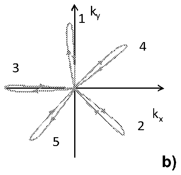
[0018]In one embodiment of the invention, the k-space sampling scheme is chosen such that it includes continuous random k-space trajectories. It is for example possible to randomly or pseudo-randomly vary a magnetic field gradient during MR signal acquisition. The k-space coordinate values as well as the time value are attributed to each MR signal sample during the acquisition procedure in order to enable reconstruction. The technical requirements for the gradient system of the used MR apparatus are low with this embodiment of the invention. No particularly fast gradient switching is required. The noise level caused by the gradient switching can be kept well within tolerable limits for the examined patient.

Problems solved by technology

However, the speed at which the MR signal data required for reconstruction of a MR image can be collected is fundamentally limited by physical and physiological constraints.

Method used


Image

  • MR image reconstruction using compressed sensing
  • MR image reconstruction using compressed sensing
  • MR image reconstruction using compressed sensing

Examples


Embodiment Construction

[0034]With reference to FIG. 1, a MR device 1 is shown. The device comprises superconducting or resistive main magnet coils 2 such that a substantially uniform, temporally constant main magnetic field is created along a z-axis through an examination volume.

[0035]A magnetic resonance generation and manipulation system applies a series of RF pulses and switched magnetic field gradients to invert or excite nuclear magnetic spins, induce magnetic resonance, refocus magnetic resonance, manipulate magnetic resonance, spatially and otherwise encode the magnetic resonance, saturate spins, and the like to perform MR imaging.

[0036]More specifically, a gradient pulse amplifier 3 applies current pulses to selected ones of gradient coils 4, 5 and 6 along x, y and z-axes of the examination volume. A digital RF frequency transmitter 7 transmits RF pulses or pulse packets, via a send- / receive switch 8, to a whole-body volume RF coil 9 to transmit RF pulses into the examination volume. A typical MR ...


PUM


Abstract

The invention relates to a method of MR imaging of an object (10) placed in an examination volume of a MR device (1). The method comprises the steps of: subjecting the object (10) to an imaging sequence for acquiring MR signal data, wherein the MR signal data are acquired as a function of k-space position and time by using an irregular k-space sampling pattern with sub-sampling of k-space; reconstructing MR image data from the MR signal data, which MR image data comprise spatial dimensions and a frequency dimension, sparsity of the MR image data in a transform domain being exploited for suppressing sub-sampling artefacts in the MR image data. Moreover, the invention relates to a MR device (1) and to a computer program.

Description

CROSS REFERENCE TO PRIOR APPLICATIONS[0001]This application is the U.S. National Phase application under 35 U.S.C. § 371 of International Application No. PCT / IB2014 / 059660, filed on Mar. 12, 2014, which claims the benefit of U.S. Patent Application No. 61 / 803,929, filed on Mar. 21, 2013. These applications are hereby incorporated by reference herein.FIELD OF THE INVENTION[0002]The invention relates to the field of magnetic resonance (MR) imaging. It concerns a method of MR imaging of at least a portion of a body of a patient placed in an examination volume of a MR device. The invention also relates to a MR device and to a computer program to be run on a MR device.BACKGROUND OF THE INVENTION[0003]Image-forming MR methods which utilize the interaction between magnetic fields and nuclear spins in order to form two-dimensional or three-dimensional images are widely used nowadays, notably in the field of medical diagnostics, because for the imaging of soft tissue they are superior to oth...

Claims


Application Information

Patent Timeline
Patent Type & Authority Patents(United States)
IPC IPC(8): G01R33/48G01R33/561G01R33/565
CPCG01R33/4824G01R33/561G01R33/56545G01R33/5611G01R33/4828
Inventor FUDERER, MIHADONEVA, MARIYA IVANOVA
Owner KONINKLILJKE PHILIPS NV
Why Eureka
Try Eureka