Computed tomography enhanced fluoroscopic system, device, and method of utilizing the same

a computed tomography and fluoroscopic system technology, applied in tomography, catheters, applications, etc., can solve the problems of difficult identification with conventional fluoroscopy, affecting the accuracy of clinical care, and requiring clinicians to provide all the information

Active Publication Date: 2018-05-22
COVIDIEN LP
View PDF191 Cites 16 Cited by
  • Summary
  • Abstract
  • Description
  • Claims
  • Application Information

AI Technical Summary

Benefits of technology

[0010]As can be appreciated, a microwave ablation catheter that is positionable through one or more branched luminal networks of a patient to treat tissue may prove useful in the surgical arena.
[0012]According to one aspect of the present disclosure, a method of enhanced navigation is provided including planning a navigation path to a target using a first data set of computed tomography images previously acquired, navigating a marker placement device to the target using the navigation path, placing a plurality of markers in tissue proximate the target, acquiring a second data set of computed tomography images including the plurality of markers, planning a second navigation path to a second target using the second data set of computed tomography images, navigating a medical instrument to the second target; capturing fluoroscopic data of tissue proximate the markers, and registering the fluoroscopic data to the second data set of computed tomography images based on marker position and / or orientation within the fluoroscopic data and the marker position and / or orientation within the second data set of computed tomography images.
[0017]Additionally, the computing device may further enable navigation of a microwave ablation device to the target and activation of the microwave ablation device to ablate tissue proximate the target. Additionally, the computing device may further analyze the fluoroscopic data and determine whether device medical instrument is correctly positioned relative to the target. A second fluoroscopic data set of the first or second target may also be acquired from a second perspective relative to the patient such that a three-dimensional position of the medical instrument is viewable from a different angle. The second fluoroscopic data set may also be analyzed to determine whether the three-dimensional position of the medical instrument relative to the target tissue is correct, and if not, the three-dimensional position of the medical instrument relative to the target may be adjusted.

Problems solved by technology

Although medical instruments like catheters, biopsy tools, etc., are clearly visible on a fluoroscopic picture, organic features such as soft tissue, blood vessels, suspicious tumor lesions etc., are either somewhat or completely transparent and thus hard to identify with conventional fluoroscopy.
However, although the medical instrument is visible in the fluoroscopic image, the area of interest or target tissue is generally somewhat transparent and not necessarily clearly visible within the image.
Moreover, fluoroscopic images render flat 2D images on which it can be somewhat challenging to assess three-dimensional position of the medical instrument.
As such, the clinician is not provided all the information that could be desired to visualize the placement of the medical device within the patient's body relative to the area of interest.

Method used


Image

  • Computed tomography enhanced fluoroscopic system, device, and method of utilizing the same
  • Computed tomography enhanced fluoroscopic system, device, and method of utilizing the same
  • Computed tomography enhanced fluoroscopic system, device, and method of utilizing the same

Examples


Embodiment Construction

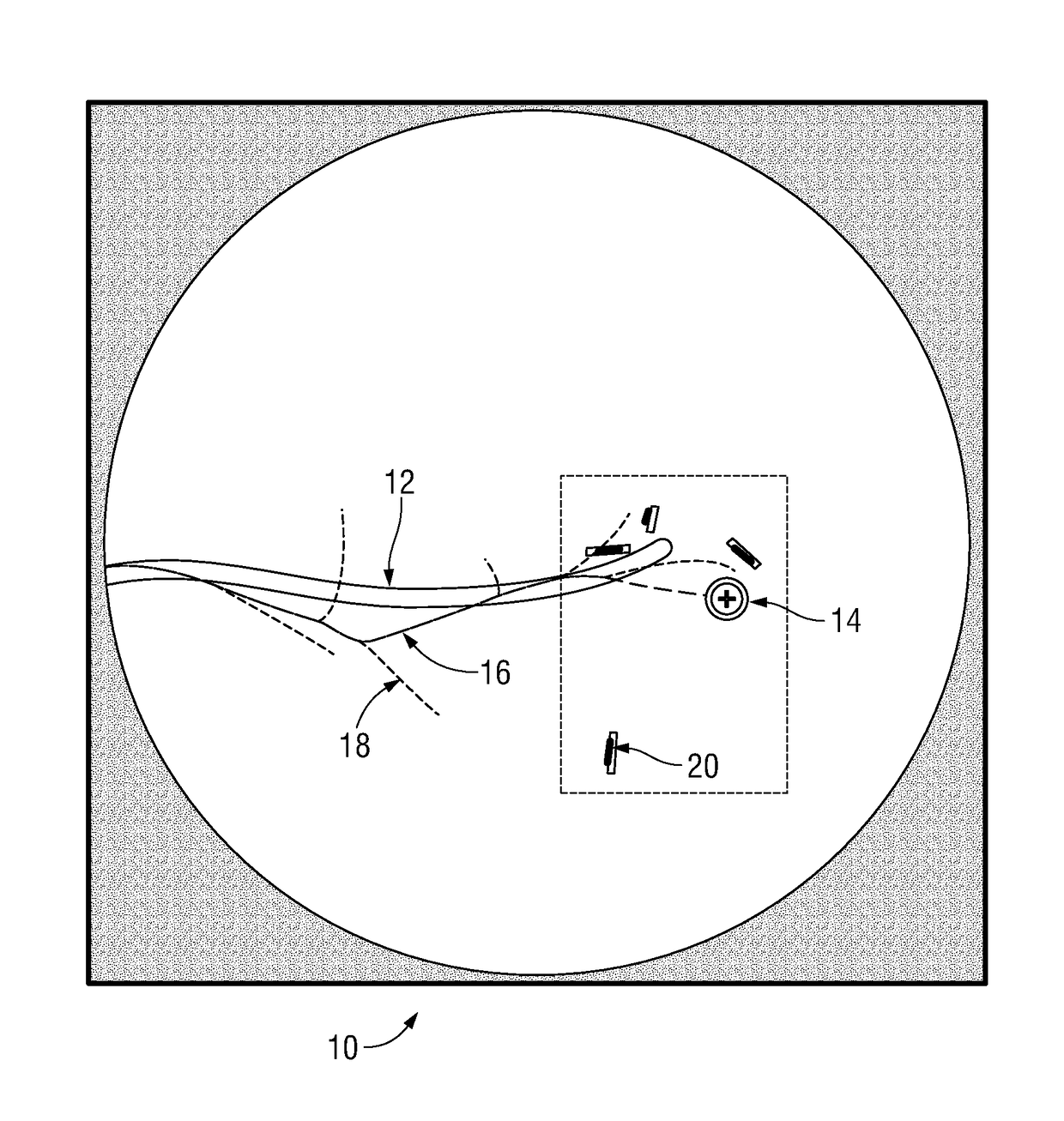
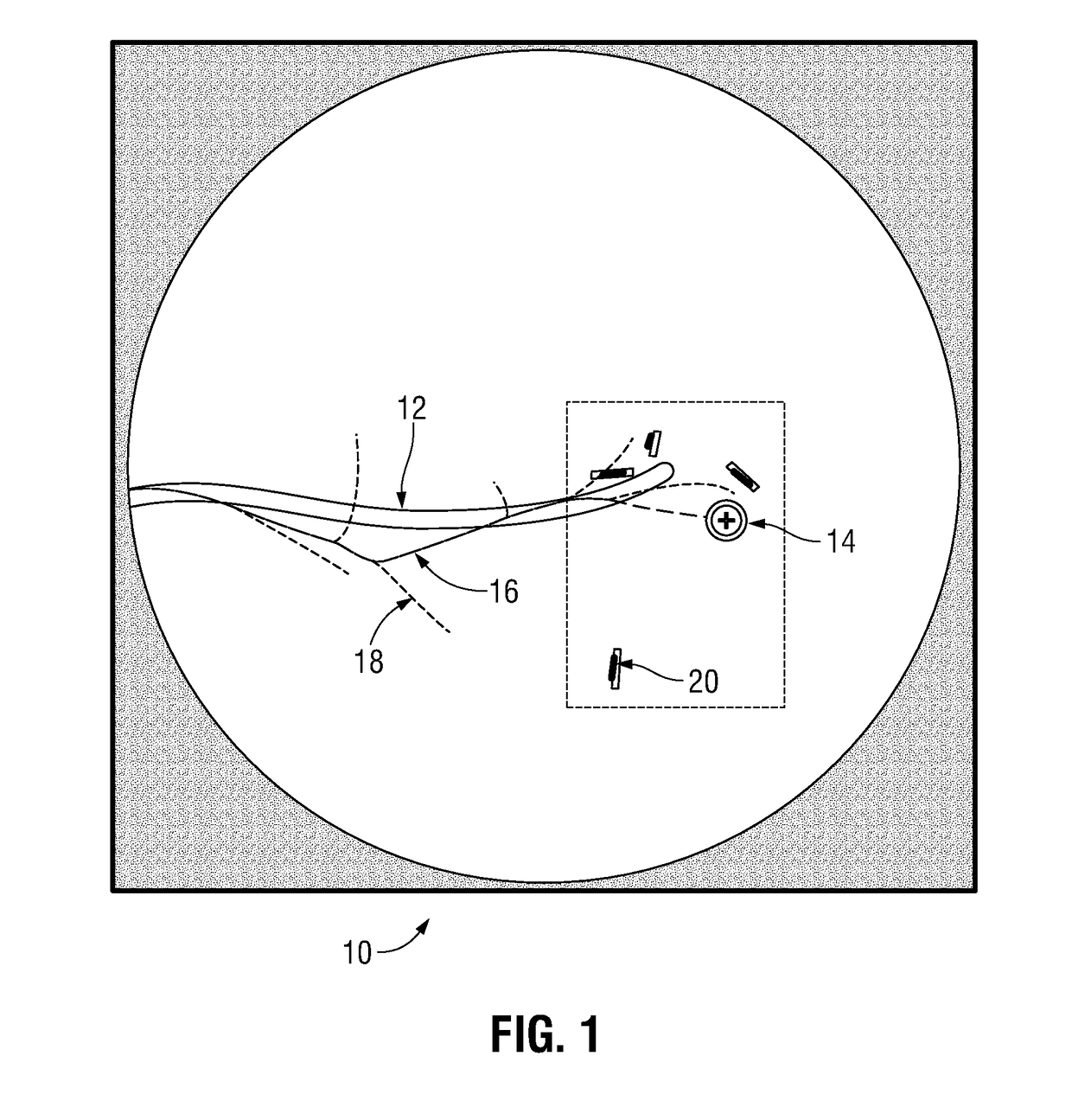
[0026]The present disclosure is generally directed to addressing the navigational and location confirmatory shortcomings of the previously known navigation and fluoroscopic imaging confirmation methods and devices. According to one embodiment of the present disclosure, following navigation of a catheter to an area of interest, a fluoroscopic image (or series of fluoroscopic images) is captured. By registering the location of markers previously placed within the patient and captured in the fluoroscopic image to the location of markers which appear in 3D model data generated from a previously acquired CT image data set, the fluoroscopic image can be overlaid with data from the 3D model data including target location data, navigation pathway data, luminal network data and more.

[0027]Detailed embodiments of the present disclosure are disclosed herein. However, the disclosed embodiments are merely examples of the disclosure, which may be embodied in various forms and aspects. Therefore, ...


PUM


Abstract

A system and method for enhanced navigation for use during a surgical procedure including planning a navigation path to a target using a first data set of computed tomography images previously acquired; navigating a marker placement device to the target using the navigation path; placing a plurality of markers in tissue proximate the target; acquiring a second data set of computed tomography images including the plurality of markers; planning a second navigation path to a second target using the second data set of computed tomography images; navigating a medical instrument to a second target; capturing fluoroscopic data of tissue proximate the target; and registering the fluoroscopic data to the second data set of computed tomography images based on marker position and orientation within the real-time fluoroscopic data and the second data set of computed tomography images.

Description

CROSS REFERENCE TO RELATED APPLICATION[0001]The present application claims the benefit of and priority to U.S. Provisional Application Ser. Nos. 62 / 073,287 and 62 / 073,306, filed on Oct. 31, 2014. This application is related to U.S. patent application Ser. No. 14 / 880,361, filed on Oct. 12, 2015. The entire contents of each of the above applications are hereby incorporated herein by reference.BACKGROUND[0002]Technical Field[0003]The present disclosure relates to a system, apparatus, and method of navigation and position confirmation for surgical procedures. More particularly, the present disclosure relates to a system and method for enhanced navigation of an extended working channel or catheter and one or more medical instruments positionable therethrough in one or more branched luminal networks of a patient and confirming placement of those medical instruments prior to initiating treatment or biopsy.[0004]Description of Related Art[0005]Microwave ablation is a commonly applied method...

Claims


Application Information

Patent Timeline
Patent Type & Authority Patents(United States)
IPC IPC(8): A61B10/04A61B6/03A61B6/12A61B6/00A61B18/00A61B18/18
CPCA61B10/04A61B6/03A61B6/12A61B6/463A61B6/5235A61B6/487A61B2018/1861A61B6/461A61B6/58A61B2018/00577A61B6/032A61B2090/364A61B2090/376A61B2034/107A61B34/20A61B2034/2051A61B2090/3966A61B2090/3987A61B34/10A61B2090/3762A61B6/022
Inventor WEINGARTEN, OREN P.AVERBUCH, DORIAN
Owner COVIDIEN LP
Why Eureka
Try Eureka