
Liver segment positioning catheter

A catheter and catheter technology, applied in the field of hepatobiliary surgery, can solve the problems of difficulty in success, difficulty in manipulation, and the range of reverse diffusion staining of methylene blue

Inactive Publication Date: 2009-04-01
THE FIRST AFFILIATED HOSPITAL OF THIRD MILITARY MEDICAL UNIVERSITY OF PLA
View PDF8 Cites 0 Cited by
  • Summary
  • Abstract
  • Description
  • Claims
  • Application Information

AI Technical Summary

Problems solved by technology

The disadvantage is that the operation is difficult, and the operator needs to be familiar with and proficient in ultrasound technology and puncture knowledge
However, for surgeons, unless they are trained accordingly, the puncture is generally not easy to succeed, and in practice, even if the puncture is successful, it is easy to cause the reverse diffusion of methylene blue to expand the staining range or quickly excrete, and the boundary disappears; There is a risk of needle tract spread
[0008] 4) Follow the method of hepatic lobectomy, such as dissecting the first hepatic hilum, freeing the left and right hepatic hilums, and blocking them separately. This method can only determine the right anterior and right posterior blood vessels and block or cut them off, and cannot reach the branches of the liver segment, which is easy Causes too much healthy liver destruction
The existing resection instruments can fully meet the requirements of liver segment resection, but the judgment technology of liver segment has the disadvantages of poor judgment accuracy and / or high technical difficulty, difficult to control, and even the risk of worsening the disease

Method used


Image

  • Liver segment positioning catheter
  • Liver segment positioning catheter
  • Liver segment positioning catheter

Examples


Embodiment 1

[0023] Example 1: Determination of the 7th segment of the liver, which is suitable for 7-segment resection

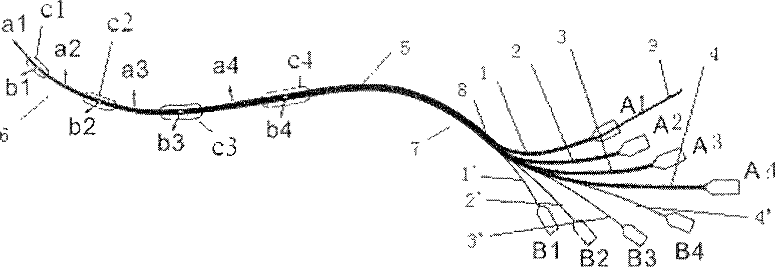
[0024] 1. After the operation begins, perform distal ligation through the right gastroepiploic vein, insert the guide wire 9 into the catheter 1, and under the guidance of the guide wire 9, place the balloon unit of the liver segment positioning catheter from the proximal end. entry vein;

[0025] 2. The operator guides the balloon unit into the right or left portal vein branch by hand;

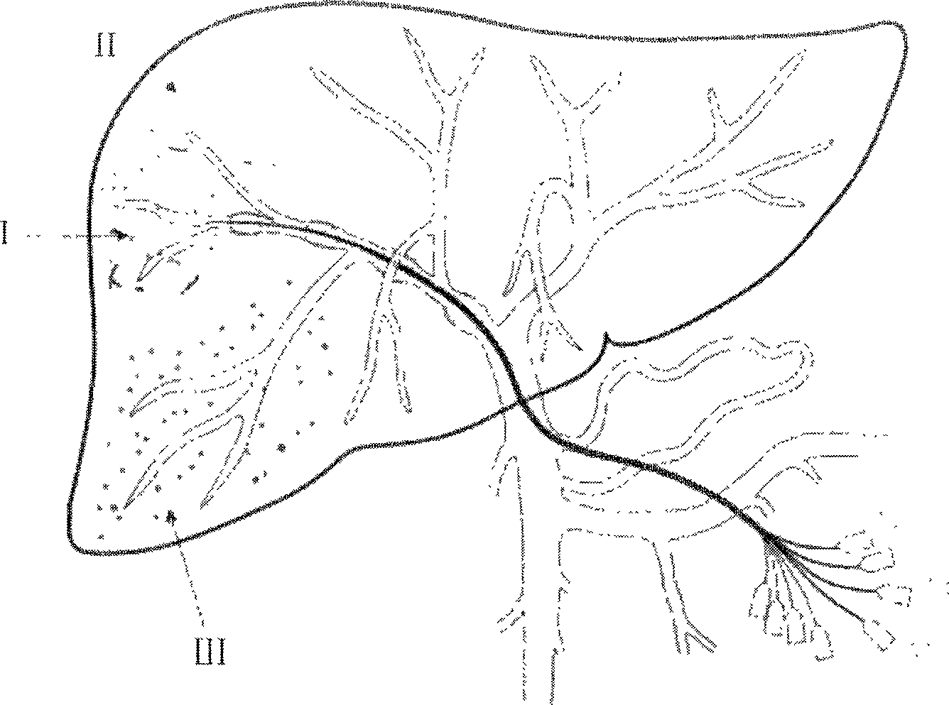
[0026] 3. After the balloon unit is placed in the right portal vein, it is continuously fed to the end of the portal vein, and the front end airbag c1 is inflated through the gas injection port B1. At this time, it is very simple to use B-ultrasound to detect on the surface of the liver. It is obtained whether the position of the balloon unit is the right anterior lobe of the liver or the right posterior lobe of the liver;

[0027] 4. If image 3 As shown, the balloon unit is assume...

Embodiment 2

[0031] Example 2 Determination of the fifth segment of the liver

[0032] 1. The liver segment positioning catheter is located in the right anterior lobe of the liver for the first time, and the positioning of the fifth and eighth segment of the liver is the same as the seventh segment in the above-mentioned embodiment 1, and the determination steps are the same;

[0033] 2. If the assumptions in Example 1 are continued, and the first liver segment positioning catheter is positioned in the right posterior lobe liver, the following steps are performed:

[0034] ①After inflating through the air supply port B1, the air supply port B2, the air supply port B3 and the air supply port B4 in sequence, and then in turn through the liquid supply port A1, the liquid supply port A2, the liquid supply port A3 and the liquid supply port A4, respectively inject Gradual dilution of melanin until the right anterior staining appears, then it is determined that the outlet ax is the channel leadi...


PUM


Abstract

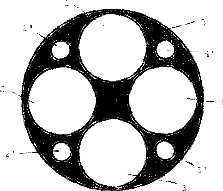
The invention relates to a liver positioning tube, formed by main body and guide wire made from macromolecule materials, wherein the front of main body has at least three ball bubbles; each ball bubble has liquid outlet of liquid tube, air outlet of air tube and air bubble; the liquid tube and air tube are in the main body; the liquid outlet and air outlet extend out the front tube of main body distantly; the air bubble through to the air inlet is on the outer tube of main body; the liquid inlet and the air inlet extend outside the main body; the wire is longer than liquid tube; the wire is smaller then liquid tube. The inventive tube can use multi-chamber multi-bubble to judge the integral interface of liver, to mark target liver in short time.

Description

technical field [0001] The invention belongs to the field of hepatobiliary surgery and relates to a catheter for positioning liver segments in hepatectomy. Background technique [0002] With the advancement of surgical technology and theory, the level of liver surgery has also been greatly improved. Diseases that were incurable in the past have been cured, and unresectable liver cancer can now be resected. Advances in surgical instruments, such as the clinical application of ultrasonic suction knife Cusa, bloodless scalpel TissueLink, etc., have led to the rapid development of liver resection technology, and doctors can perform resection with ease. With resection, there is less blood loss during the operation, minimal disturbance to the pathophysiology of the patient, and the operation can be passed safely and smoothly. The patient's economic affordability and perioperative management skills have been greatly improved compared with the past, which provide greater support fo...

Claims


Application Information

Patent Timeline
Patent Type & Authority Patents(China)
IPC IPC(8): A61M25/00A61M39/08A61B19/00A61B17/00A61B90/00A61M25/10
Inventor 冯晓彬
Owner THE FIRST AFFILIATED HOSPITAL OF THIRD MILITARY MEDICAL UNIVERSITY OF PLA
Learn More