Fault image segmentation method for images scanned from films

A technology of tomographic images and scanned images is applied in the field of segmentation of tomographic images of CT film scanning images, which can solve the problems of low efficiency and poor effect, and achieve the effects of high applicability, good consistency and improved positioning accuracy.

Inactive Publication Date: 2007-07-25
SICHUAN UNIV
View PDF0 Cites 11 Cited by
  • Summary
  • Abstract
  • Description
  • Claims
  • Application Information

AI Technical Summary

Problems solved by technology

This method is low in efficiency and poor in effect because it h

Method used


Image

  • Fault image segmentation method for images scanned from films
  • Fault image segmentation method for images scanned from films
  • Fault image segmentation method for images scanned from films

Examples


Example Embodiment

[0018] In the following, in conjunction with the accompanying drawings, the method for segmenting the interrupted layer image of the CT film scan image of the present invention will be further explained in detail.

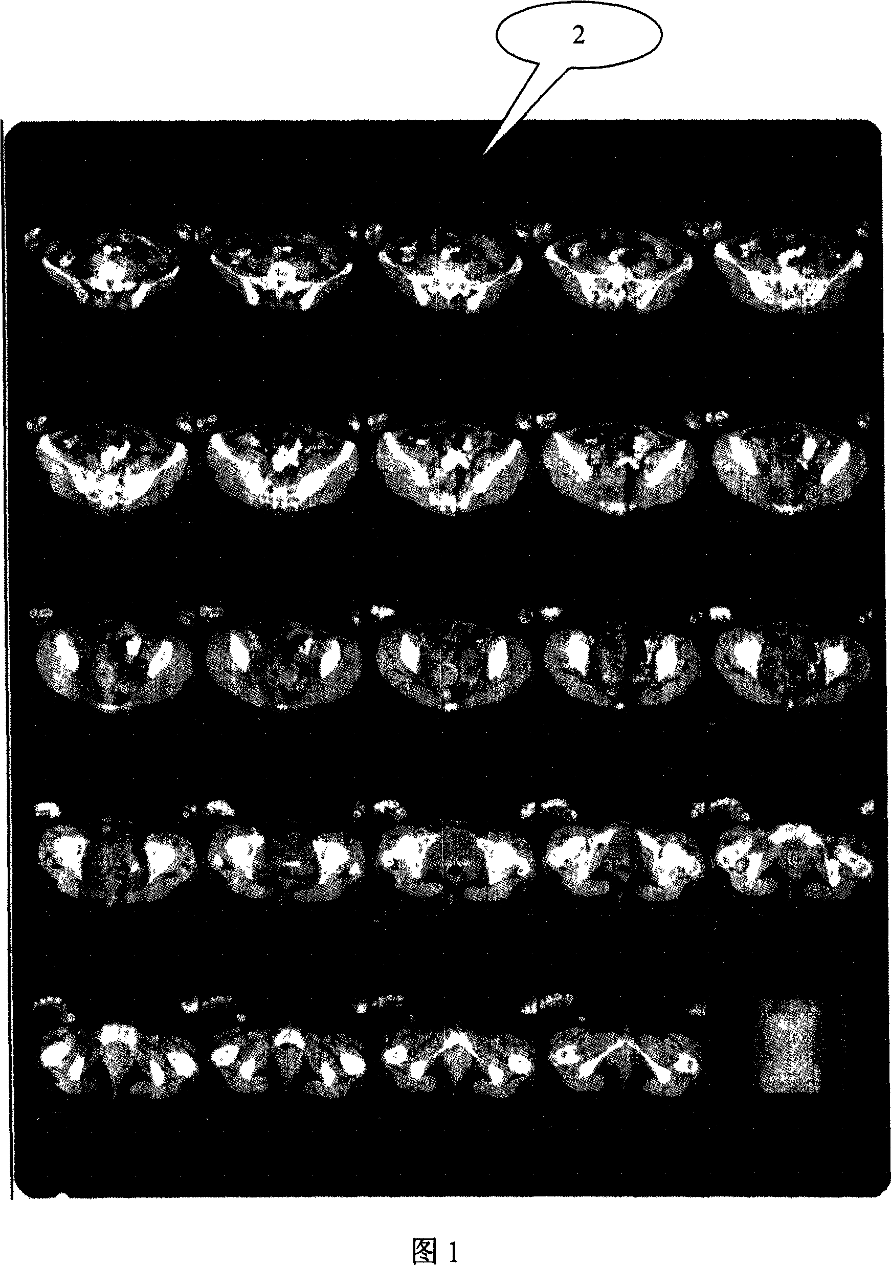
[0019] Figure 1 is a CT film scan image to be segmented. In the figure, there are several CT tomographic images arranged on a CT film. Therefore, before the 3D reconstruction, the tomographic image must be segmented from the film scan image one by one.

[0020] In this embodiment, the specific steps of the tomographic image segmentation method are:

[0021] 1) Determine the resolution of the reference tomographic image according to the length actually represented by the scale and the pixel coordinates of the two endpoints on the scanned image;

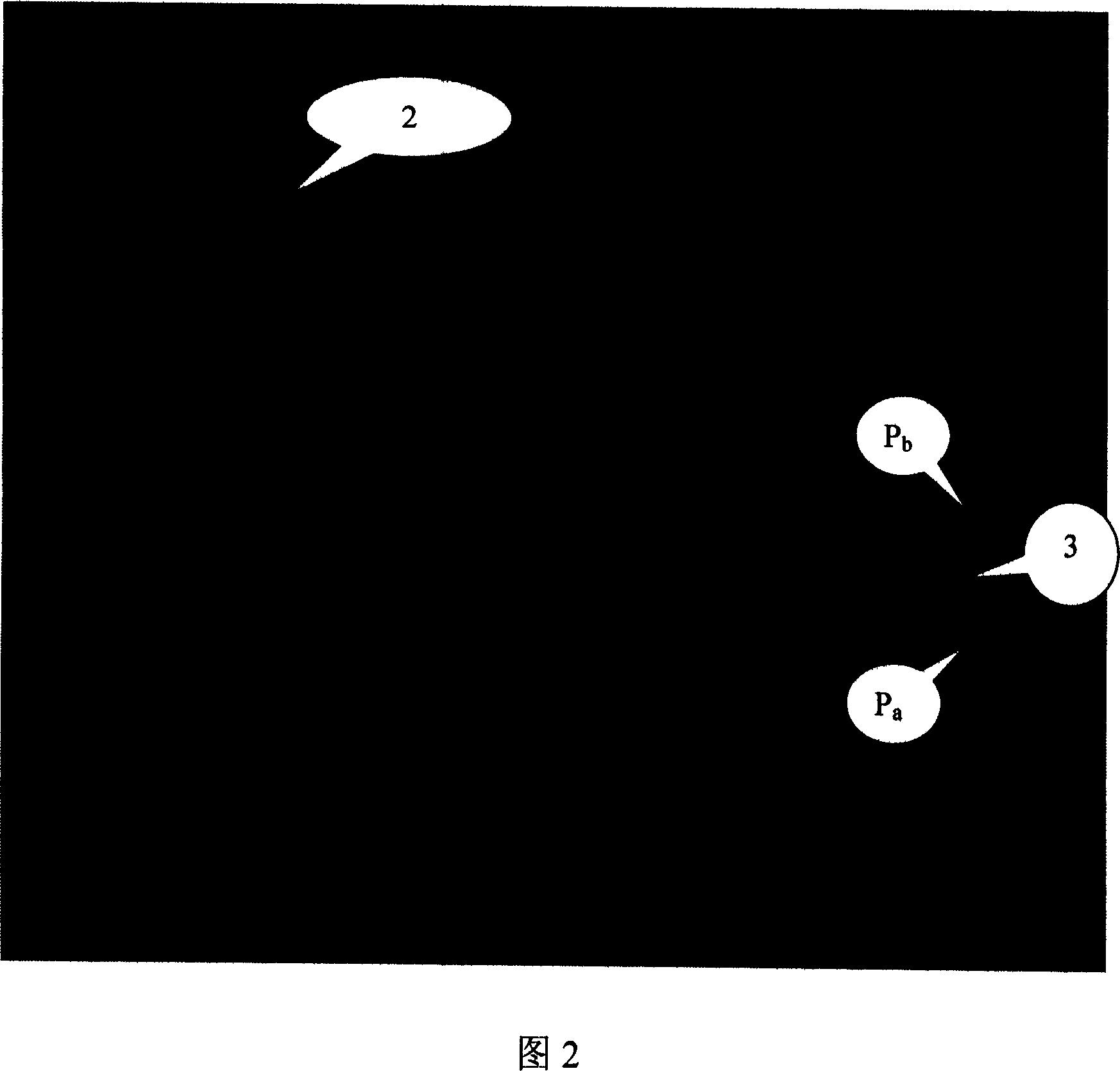
[0022] The CT film scan is made into a CT film scan image, and one of the tomographic images that need to be segmented is used as a reference image, and the two end points of the reference tomographic image ruler are automatica...


PUM


Abstract

A method for dividing tomograph in scanned image of CT film includes confirming resolution of reference tomography, selecting tomograph to be divided, using scale end point of reference tomography as reference point, locking relative position relation of rectangular frame to scale reference point, conforming resolution of the other tomography and carrying out convergent-divergent on the other tomograph as per set ratio, using scale end point of the other tomography as reference to confirm rectangular frame position in the other tomograph and using region surrounded by said frame as tomograph to be divided.

Description

technical field [0001] The invention relates to medical image processing, in particular to a method for segmenting slice images of CT film scan images. Background technique [0002] In some hospitals, digitizing CT film images through film scanners is the only way to obtain human anatomical information. Usually, several CT tomographic images are arranged on one film. Therefore, before 3D reconstruction, the tomographic images must first be segmented from the film scan images one by one. In order to facilitate processing such as 3D reconstruction, software systems based on medical images usually require medical images to be parallel tomographic image sequences, and also require that the tomographic image sequences must be consistent in scale, angle, and position. [0003] In existing solutions, the consistency of the angular relationship is achieved by rotating the film-scanned image. This requires that the angles of the tomographic images on the same CT film must not devi...

Claims


Application Information

Patent Timeline
IPC IPC(8): G06T5/00A61B6/03
Inventor 吴章文罗正明勾成俊杨代伦
Owner SICHUAN UNIV
Why Eureka
Try Eureka