Device and method for isolating cells, bioparticles and/or molecules used for animal biotechnology (including biological research) and medical diagnosis from liquids
A technology of biotechnology and biological particles, applied in the direction of scientific instruments, instruments, biological tests, etc., can solve problems such as unsustainable operation
- Summary
- Abstract
- Description
- Claims
- Application Information
AI Technical Summary
Problems solved by technology
Method used
Image
Examples
Embodiment approach 1
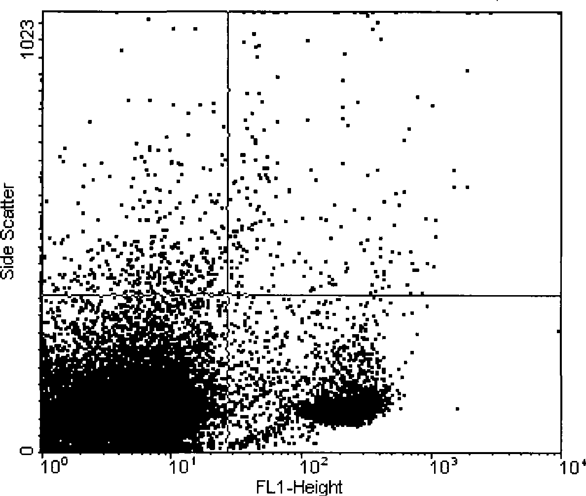
[0026] Isolation of CD4 Positive Cells from Rat Whole Blood
[0027] Ascites antibody (RIB5 / 2) was done according to the protocol of Millipore Montage's Antibody Purification Kit (LSK2ABG20).
[0028] After purification, use SDS denaturing gel (10%) to detect
[0029] Conjugation of Anti-CD4 Antibody to Polypropylene (PMA) Particles
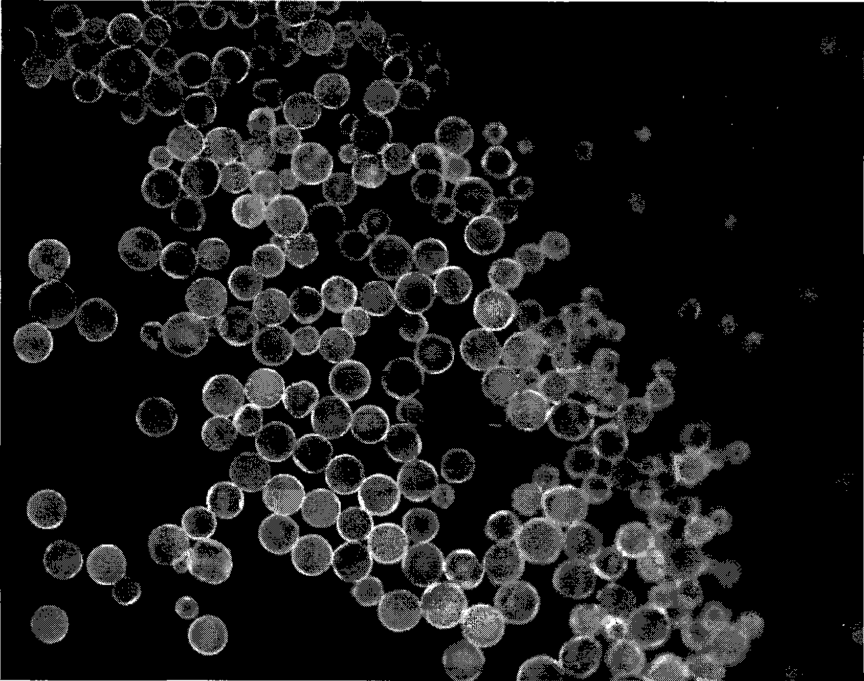
[0030] 1. Centrifuge 1ml of PMA (particle diameter=40μm+ / -10μm; 10mg / ml; COOH / PEG-COOH modification) for 2 minutes at 3,000xg, remove the supernatant and inhale in 1ml of 0.1M MES pH6.3 solution.
[0031] 2. Dissolve 2mg of EDC and 2.4mg of N-hydroxysuccinimide in 0.5ml of 0.1M MES pH6.3 buffer solution and add to the suspension of PMA particles. Incubate for 1 hour at room temperature with rotation (activation of particles).
[0032] 3. Separation of PMA particles: Centrifuge and elute twice with 0.1M MES r pH6.3 solution.
[0033] 4. Aspirate activated PMA particles into 1 ml of 0.1M MES pH6.3 buffer solution with 100-150 μg of antibody; co...
Embodiment approach 2
[0052] Protein Isolation (IgG) from Cell Lysates
[0053] Ascites antibody (RIB5 / 2) was done according to the protocol of Millipore Montage's Antibody Purification Kit (LSK2ABG20).
[0054] Purification was then checked with SDS denaturing gel (10%).
[0055] (Mouse IgG 2) Conjugation of Anti-CD4 Antibody to Polypropylene (PMA) Particles
[0056] 1. Centrifuge 1ml of PMA (particle diameter=40μm+ / -10μm; 10mg / ml; COOH / PEG-COOH modification) for 2 minutes at 3,000xg, remove the supernatant and inhale in 1ml of 0.1M MES pH6.3 buffer solution.
[0057] 2. 2mg of EDC and 2.4mg of N-hydroxysuccinimide buffered in 0.5ml of 0.1M MES pH6.3 buffer solution were added to the suspension of PMA particles. Incubate for 1 hour at room temperature with rotation (activation of particles).
[0058] 3. Separation of PMA particles: Centrifuge and elute twice with 0.1M MES r pH6.3 buffer solution.
[0059] 4. Aspirate active PMA particles into 1ml 0.1M MES pH6.3 buffer solution with 100-150μg ...
Embodiment approach 3
[0074] Figure 7-11 shows an example of the invention or a part thereof implemented on the basis of an instrument, Figure 7 Represents a typical system that can be used continuously. The dotted arrow indicates the hose system. The arrow tip marks the flow direction of the fluid. Starting from the organism (mammal), the body fluid, such as blood, is pumped or directed to a container containing functional particles. The reaction vessel, possibly through a blood pump or a vacuum tube (marked as a circle in Figure 8), interacts in admixture with the constituents of the body fluid. directing a medium of cell mixture and functional particles to a particle separation system, according to one example, the medium of cell mixture and functional particles is directed to a sieve and passed through a hollow fiber membrane, and then separated into a cell mixture free of functional particles and a medium containing Cell mixture of functional particles ( Figure 9 ). But the mixture witho...
PUM
| Property | Measurement | Unit |
|---|---|---|
| diameter | aaaaa | aaaaa |
| diameter | aaaaa | aaaaa |
Abstract
Description
Claims
Application Information
 - R&D
- Intellectual Property
- Life Sciences
- Materials
- Tech Scout
- Unparalleled Data Quality
- Higher Quality Content
- 60% Fewer Hallucinations
Browse by: Latest US Patents, China's latest patents, Technical Efficacy Thesaurus, Application Domain, Technology Topic, Popular Technical Reports.
© 2025 PatSnap. All rights reserved.Legal|Privacy policy|Modern Slavery Act Transparency Statement|Sitemap|About US| Contact US: help@patsnap.com


