
Method of preparing containing endothelium ancestor cell preparation utilizing umbilical or placenta and uses thereof

A technology of endothelial progenitor cells and placenta, applied in biochemical equipment and methods, animal cells, vertebrate cells, etc., can solve the problems of endothelial progenitor cell sources and poor proliferation ability, and achieve good proliferation potential, abundant sources, and large-scale supply Effect

Inactive Publication Date: 2008-06-18
INST OF HEMATOLOGY & BLOOD HOSPITAL CHINESE ACAD OF MEDICAL SCI
View PDF0 Cites 13 Cited by
  • Summary
  • Abstract
  • Description
  • Claims
  • Application Information

AI Technical Summary

Problems solved by technology

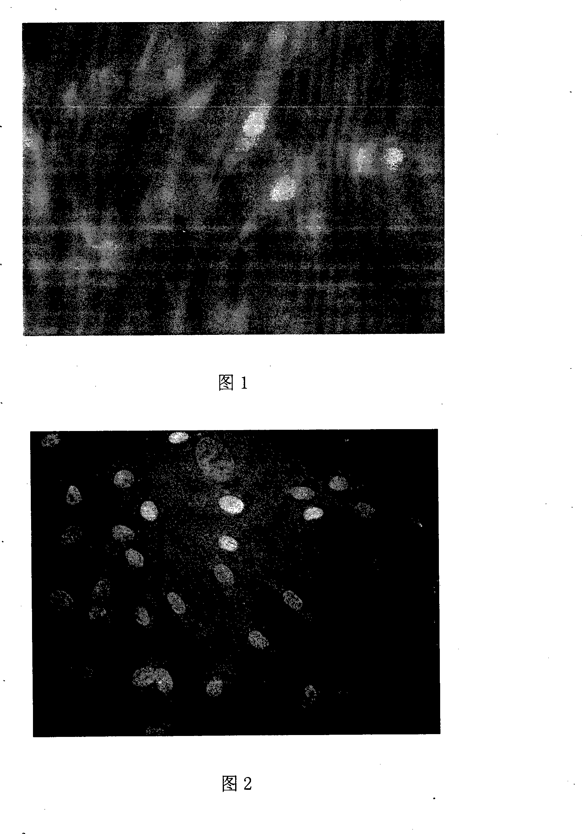
[0007] The technical problem to be solved by the present invention is to provide a method for preparing a preparation containing endothelial progenitor cells by using umbilical cord or placenta and its application, that is, a preparation method of a new source of endothelial progenitor cell preparation, in which umbilical cord and placenta-derived mesenchymal cells In the differentiation experiment, it was found that mesenchymal stem cells can easily differentiate into endothelial progenitor cells with a transformation rate of more than 80%, and they still have the ability to expand during and after transformation. The problem provides a solution

Method used


Image

  • Method of preparing  containing endothelium ancestor cell preparation utilizing umbilical or placenta and uses thereof

Examples


Embodiment 1

[0034] Example 1: Preparation of endothelial progenitor cells differentiated from umbilical cord-derived mesenchymal cells and its preparation method

[0035] A preparation of umbilical cord mesenchymal stem cells

[0036] 1. Take the umbilical cord of a healthy full-term baby and wash the surface with saline solution containing gentamicin (concentration 20,000-40,000 U / L) and heparin (concentration 50-200 U / ml) to remove residual blood and possible bacterial contamination , at least three times, each time the container and equipment are replaced.

[0037] 2. Mechanically crush the tissue, add 4-5 times the volume of collagenase digestion solution, stir at 37°C, collect the digestion solution after 2 hours, filter through a 200-mesh filter, add 2 times the volume of washing solution (DF12 plus 5- 20% fetal bovine serum) and washed twice, use mesenchymal medium (containing 10-20% fetal bovine serum, 10ng / ml bFGF, 20ng / ml EGF) to resuspend the pellet and inoculate it in a cultu...

Embodiment 2

[0052] Example 2: Preparation of endothelial progenitor cells differentiated from placenta-derived mesenchymal cells and its preparation method A Preparation of placental mesenchymal stem cells

[0053] 1. Take the placenta of a healthy full-term baby and wash the surface with saline solution containing gentamicin (concentration 20,000-40,000 U / L) and heparin (50-200 U / ml) to remove residual blood and possible bacterial contamination. At least three times, each time the container and instrument are replaced.

[0054] 2. Mechanically crush the tissue, add 4-5 times the volume of collagenase digestion solution, stir at 37°C, collect the digestion solution after 2 hours, filter through a 200-mesh filter, add 2 times the volume of washing solution (DF12 plus 5- 20% fetal bovine serum) and washed twice, use mesenchymal medium (containing 10-20% fetal bovine serum 10ng / mlbFGF 20ng / ml EGF) to resuspend the pellet and inoculate it in a culture bottle, and replace it in full after 24-7...


PUM


Abstract

The invention discloses a method which adopts umbilical cord or placenta to prepare a preparation containing endothelial progenitor cells and an application of the preparation, mainly relating to a novel source of an endothelial progenitor cell preparation and a preparation method of the endothelial progenitor cell preparation. The stem cell preparation adopts the placenta or the umbilical cord as the source of the cells; mesenchymal stem cells are firstly separated and amplified; and then VEGF is added to induce the mesenchymal stem cells to be differentiated into the endothelial progenitor cells. The endothelial progenitor cell preparation prepared according to the invention contains a large number of endothelial progenitor cells. The preparation can be applied to clinical treatment and tissue engineering, etc. The raw material sources of the preparation are rich; the success rate of the separation and the yield are high; and the proliferation ability of the cells is strong.

Description

technical field [0001] The invention relates to a biological preparation, especially a method for preparing a preparation containing endothelial progenitor cells by using umbilical cord or placenta and its application. Background technique [0002] Endothelial progenitor cells (EPCs), also known as angioblasts, are precursor cells derived from bone marrow that can circulate, proliferate and differentiate into mature endothelial cells (ECs) in vivo. In vivo EPC can settle to the ischemic site and participate in the formation of new blood vessels. The genetically modified EPC is also used for anti-angiogenic treatment of neoplastic and inflammatory diseases. Using EPC as seed cells can construct tissue engineered blood vessels in animal experiments. EPCs were seeded on biodegradable scaffolds and transplanted into the inferior vena cava of dogs. The results confirmed that EPCs differentiated into endothelial cells and smooth muscle cells in dogs. At present, EPC can be isol...

Claims


Application Information

Patent Timeline
Patent Type & Authority Applications(China)
IPC IPC(8): A61K35/50C12N5/08A61P9/10A61P29/00A61P35/00A61L27/38A61K35/51C12N5/071C12N5/0775
Inventor 邱录贵孟恒星徐燕
Owner INST OF HEMATOLOGY & BLOOD HOSPITAL CHINESE ACAD OF MEDICAL SCI
Learn More