
Real-time optoacoustic monitoring with electrophysiologic catheters

A catheter, photoacoustic technology, applied in the direction of catheter, application, medical science, etc., can solve the problems of acousto-optic data irradiation and detection limitations

Active Publication Date: 2008-08-20
BIOSENSE WEBSTER INC
View PDF8 Cites 14 Cited by
  • Summary
  • Abstract
  • Description
  • Claims
  • Application Information

AI Technical Summary

Problems solved by technology

However, the in vivo application of this technique, and especially the intracardiac and intraepicardial application, is limited in providing accurate acousto-optic data due to various factors including spatial constraints and instrument integration. are limited in terms of exposure and detection

Method used


Image

  • Real-time optoacoustic monitoring with electrophysiologic catheters
  • Real-time optoacoustic monitoring with electrophysiologic catheters
  • Real-time optoacoustic monitoring with electrophysiologic catheters

Examples


Embodiment Construction

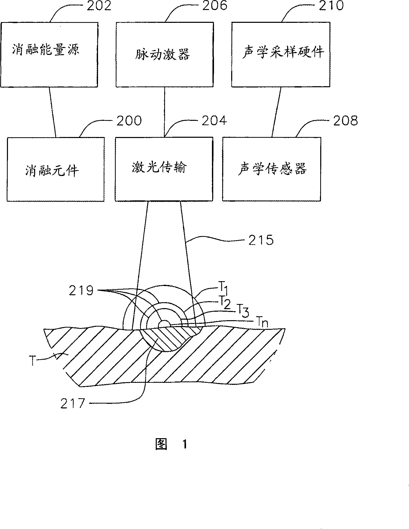
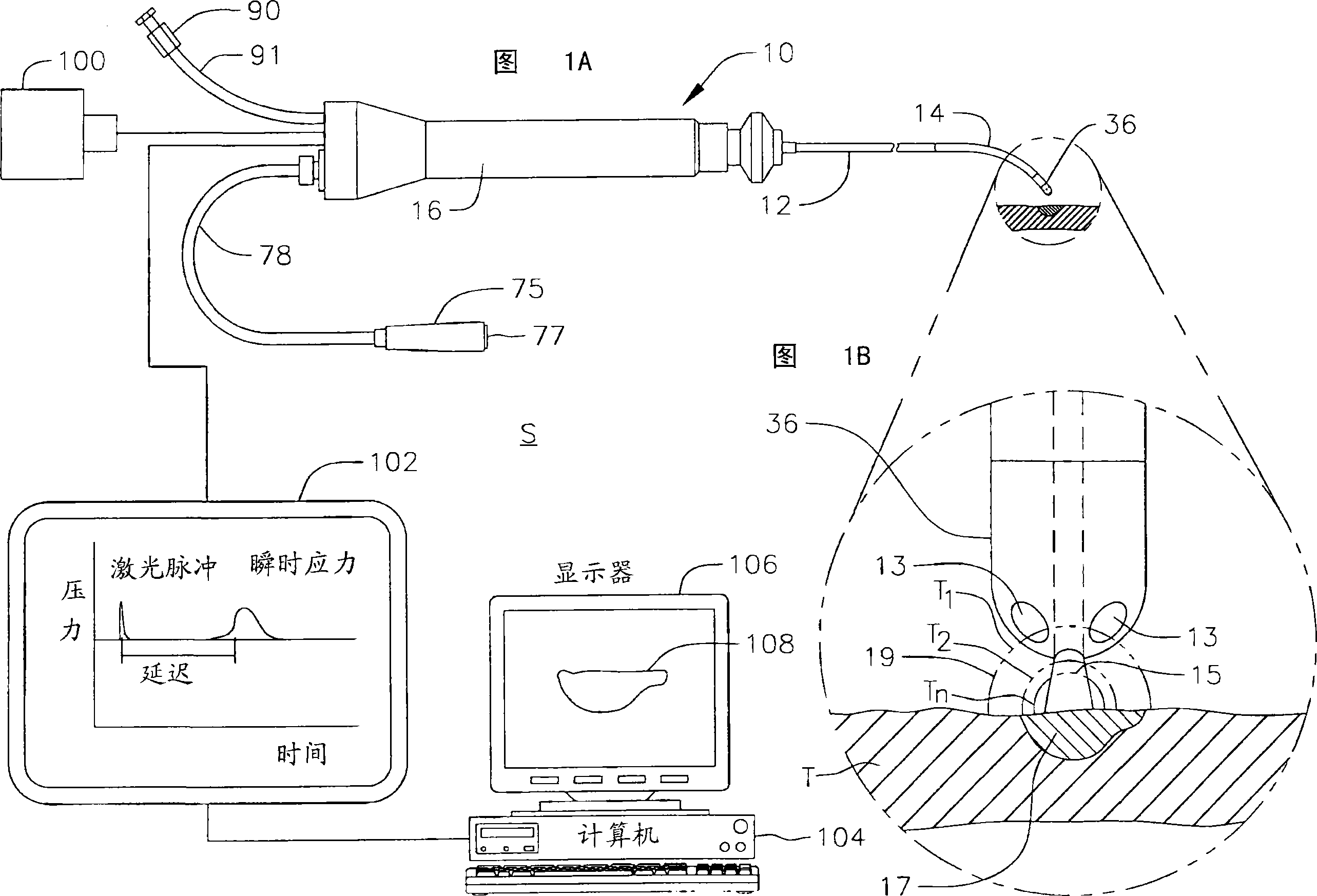
[0035]Figure 1 shows one embodiment of a system S for laser photoacoustic monitoring to provide real-time assessment of lesion formation, tissue status, and tissue morphology. Tissue T receives RF ablation from ablation element 200 powered by ablation energy source 202 to form lesion 217 . Laser delivery device 204 illuminates lesion 217 and surrounding tissue within its field of view 215 to excite pressure waves 219 (with varying delay times T1, T2 . . . Tn) that are detected by acoustic transducer 208 , used to image the injury against the background of the surrounding tissue. The laser delivery means may comprise a fiber optic cable enclosed within a conduit which is assembled separately or first for irradiation, or within an integrated conduit as further described below. As will be appreciated by those skilled in the art, the imaging provided by the present invention is based on contrast differences provided by differential absorption. To this end, the pulsating laser li...


PUM


Abstract

A system and method for opto-acoustic tissue and lesion assessment in real time on one or more of the following tissue characteristics: tissue thickness, lesion progression, lesion width, steam pop, and char formation, system includes an ablation element, laser delivery means, and an acoustic sensor. The invention involves irradiating tissue undergoing ablation treatment to create acoustic waves that have a temporal profile which can be recorded and analyzed by acoustic sampling hardware for reconstructing a cross-sectional aspect of the irradiated tissue. The ablation element (e.g., RF ablation), laser delivery means and acoustic sensor are configured to interact with a tissue surface from a common orientation; that is, these components are each generally facing the tissue surface such that the direction of irradiation and the direction of acoustic detection are generally opposite to each other, where the stress waves induced by the laser-induced heating of the tissue below the surface are reflected back to the tissue surface.

Description

technical field [0001] The present invention relates to electrophysiology catheters, and in particular to laser photoacoustic electrophysiology catheters for tissue monitoring and injury assessment. Background technique [0002] For certain types of minimally invasive medical procedures, real-time information about the condition of the treatment site within the body is difficult to obtain. This lack of information constrains the physician when performing a procedure with the catheter. An example of such procedures is the treatment of tumors and diseases in the liver and prostate. Yet another example of such a procedure is cardiac ablation for the treatment of atrial fibrillation. This condition in the heart causes abnormal electrical signals to be generated in the tissue within the heart, causing the heart to beat irregularly. [0003] Arrhythmias are most commonly caused by abnormal electrical current routing through the tissue within the heart. Typically, most arrhythm...

Claims


Application Information

Patent Timeline
Patent Type & Authority Applications(China)
IPC IPC(8): A61B5/00A61B18/00
CPCA61B5/0084A61B5/0095A61B8/12A61B18/1492
Inventor S·沙拉雷C·A·利伯尔
Owner BIOSENSE WEBSTER INC
Learn More