

Method and apparatus for sample preparation
A nucleic acid and individual technology, applied in biochemical equipment and methods, microbial measurement/inspection, fluorescence/phosphorescence, etc., can solve problems that are not suitable for digital counting, and achieve the prevention of sample loss and reduction of reaction efficiency, cost and Effects of labor saving, cost and labor reduction
- Summary
- Abstract
- Description
- Claims
- Application Information
AI Technical Summary
Problems solved by technology
Method used
Image

Examples
Embodiment 1
[0061] This example shows an example in which an emulsion containing agarose was prepared by stirring in oil.
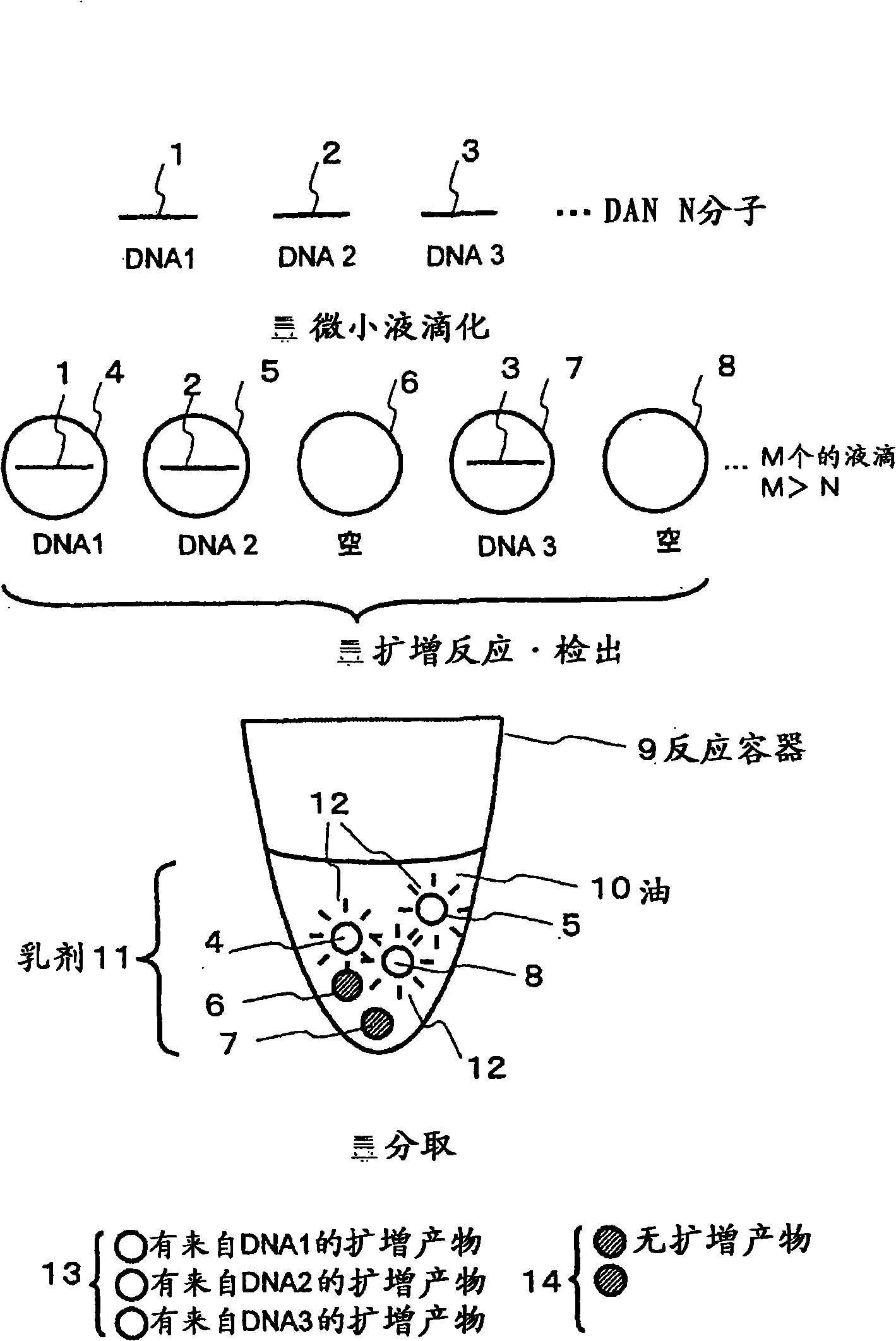
[0062] exist figure 1 The basic concept of this method is shown in . By dividing the sample solution containing DNA molecules 1-3 to be analyzed into M micro-droplets 4-8 larger than the total number N of DNA molecules, micro-droplets 4, 5, 7, DNA into which DNA has entered are formed. Tiny droplets that did not enter6,8. The tiny droplets 4-8 are dispersed in the oil 10 in the reaction vessel 9 to form an emulsion 11 . After the emulsion containing the micro-droplets is subjected to an amplification reaction such as PCR, the presence or absence (amount) of the amplification product obtained in each micro-droplet is detected by fluorescence detection using an intercalator or the like, and is divided into 12 detections with fluorescence. The micro-droplets 13 of the amplified products from each DNA 1-3 and the micro-droplets 14 without the amplified products that c...
Embodiment 2
[0100] Embodiment 2: the shape of reaction vessel
[0101] In this embodiment, the shape of the reaction vessel is formed on the plate of the series of micro reaction wells separated from each other. use Figure 8-1 0 explains this embodiment. elephant Figure 8 Likewise, the plate 80 is provided with a plurality of wells 83 for collecting the respective minute droplets 81, 82. hole 83 as Figure 9 As shown, the 2-dimensional sequence constitutes the plate 80 . The tiny droplet directly enters the hole 83 and can be covered with a hydrophobic solvent 84 or the like, or can enter the hydrophobic solvent 84 in the hole 83 .
[0102] In this case, the hydrophobic solvent 84, in addition to the purpose of forming an emulsion, can also realize the function of preventing the evaporation of water in the reaction solution, keeping the shape of the tiny droplet as a spherical function, and preventing the gel and the surface of the container when the gel is taken out. The bonding ...
Embodiment 3
[0108] This example shows another example of the method for producing fine liquid droplets.
[0109] use Figure 11 This embodiment will be described. In this embodiment, the inkjet unit 100 is used to form minute droplets. The inkjet unit 100 is composed of a tank 101 for storing a solution for preparing fine liquid droplets 103 and a nozzle 102 for forming fine liquid droplets and ejecting them. By heating the reaction liquid in the nozzle instantaneously, a certain amount of the reaction liquid is ejected. The tiny liquid droplets 103 are arranged facing the container 105 so that they are sprayed or dropped directly into the hydrophobic solvent 104 . Micro-droplets 103 prepare emulsion 106 by spraying or falling into hydrophobic solvent 104 .
[0110] This embodiment is suitable for controlling the size and quantity of tiny liquid droplets, and is especially suitable for preparing liquid droplets from about 0.5 pl to about 10 pl (about 10 μm-30 μm in diameter). It is e...
PUM
| Property | Measurement | Unit |
|---|---|---|
| diameter | aaaaa | aaaaa |
| diameter | aaaaa | aaaaa |
Abstract
Description
Claims
Application Information