Novel ultrasound medical imaging method

A technology of medical imaging and ultrasound, applied in the direction of echo tomography, etc., can solve the problem of X-ray damage to the human body, and achieve the effect of low cost

Inactive Publication Date: 2008-12-10
徐州雷奥医疗设备有限公司
View PDF0 Cites 9 Cited by
  • Summary
  • Abstract
  • Description
  • Claims
  • Application Information

AI Technical Summary

Problems solved by technology

The traditional ultrasonic medical imaging uses the reflection method, that is, an ultrasonic probe is used to transmit and then receive under the external excitation signal for scanning tomography. At the end of the 20th century, it was used for X-ray scanning tomography of the head and the whole body. The advent of imaging (X-CT) has created a new chapter of epoch-making significance for medical diagnosis, but X-rays are harmful to the human body and require ultra-high voltage power supply

Method used


Image

  • Novel ultrasound medical imaging method
  • Novel ultrasound medical imaging method
  • Novel ultrasound medical imaging method

Examples


Embodiment Construction

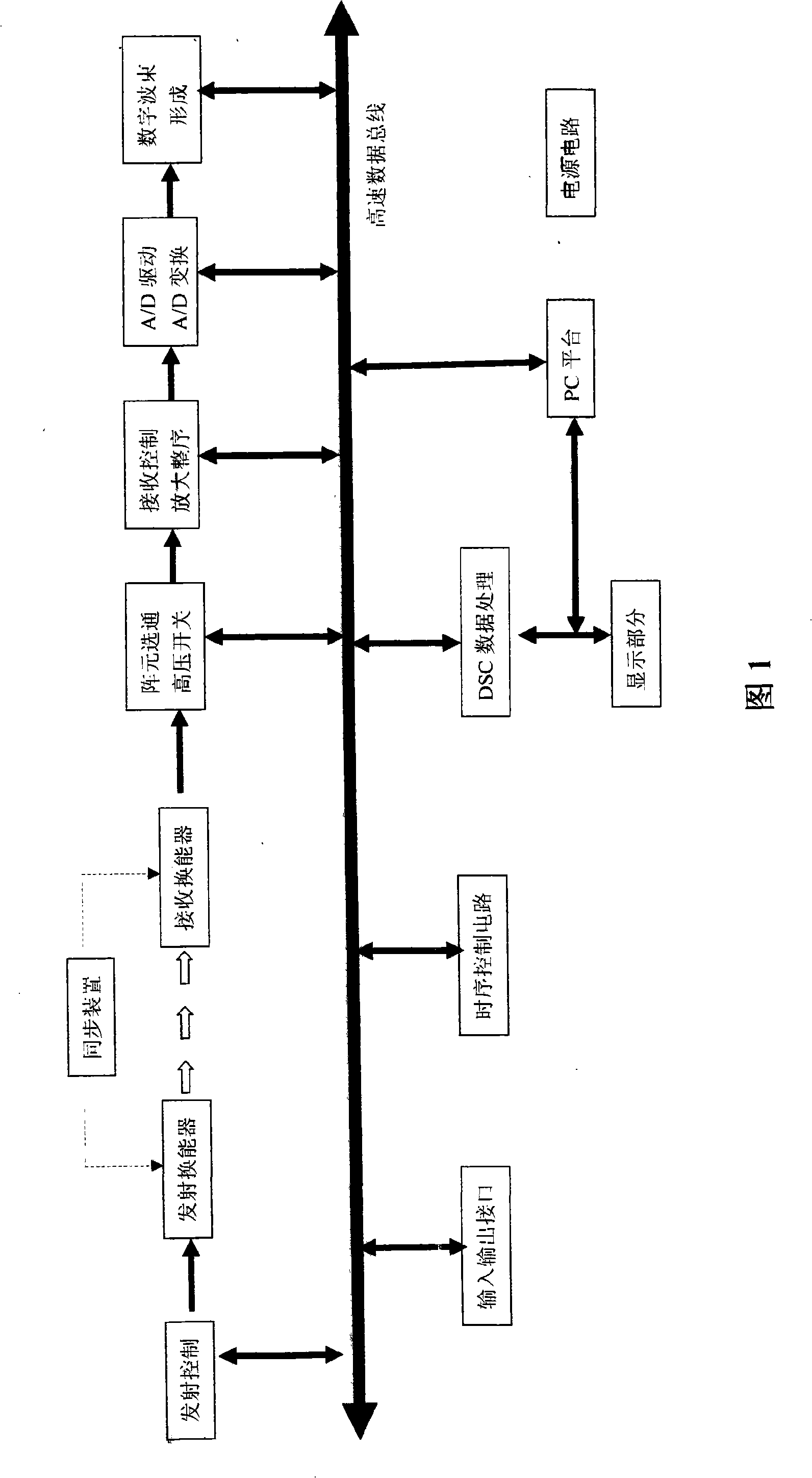
[0032] Referring to the system composition block diagram shown in Fig. 1, it indicates the signal flow between each functional module and the control unit in the device of the present invention. It is a full-digital ultrasonic medical imaging device based on a PC platform. The specific device includes a synchronization device, a transmitting control part, an ultrasonic transmitting transducer, an ultrasonic receiving transducer, an array element gating high-voltage control part, and a receiving control amplification sequence part. , A / D drive and conversion part, digital beam forming part, DSC data processing part, display unit, PC platform, power supply unit and other parts.

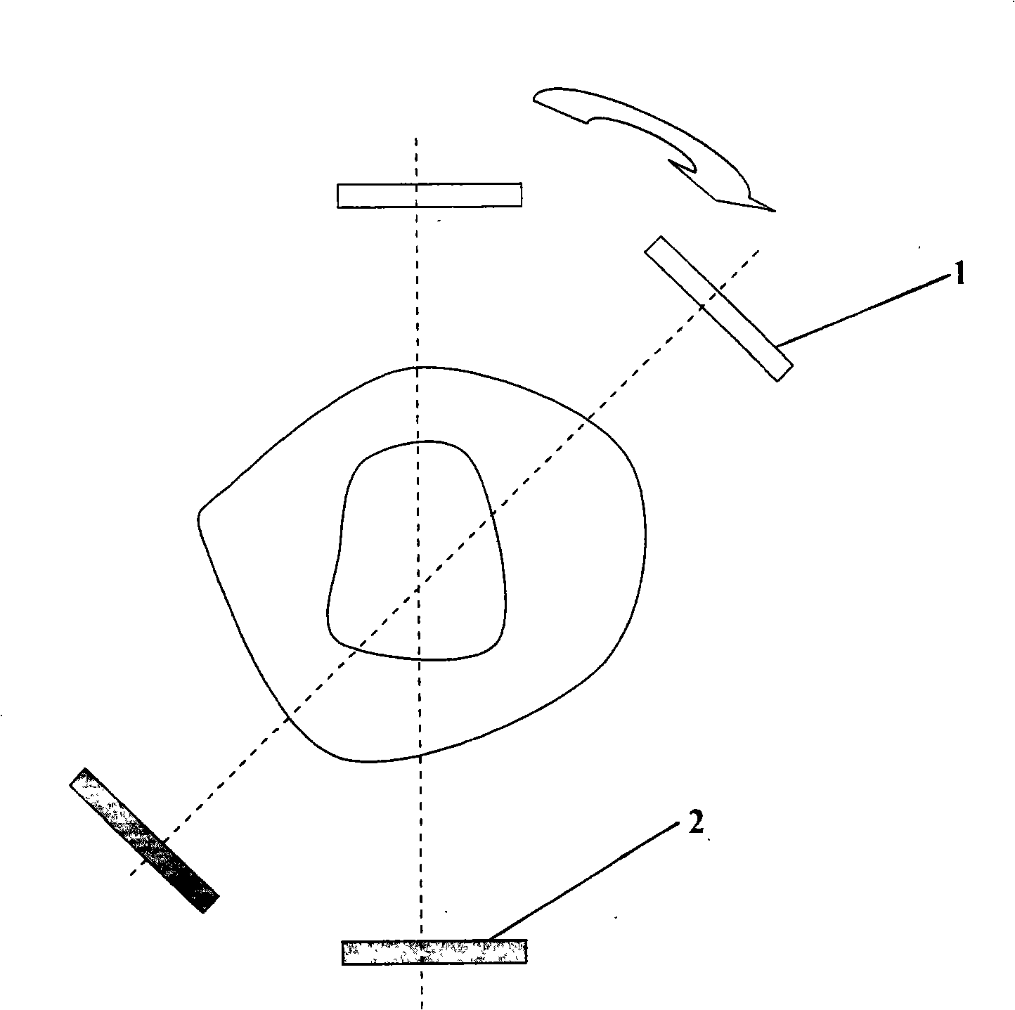
[0033] refer to figure 2 As shown, it can make the coaxial transmitting transducer and receiving transducer scan along a straight line to obtain the projection data in the cut plane, and then the pair of transmitting and receiving transducers rotate an angle in the same plane, and then Scanning in a s...


PUM


Abstract

The invention discloses an ultrasonic medical imaging method by utilizing a transmission method. As a novel ultrasonic medical imaging method, the invention is a fully digitalized ultrasonic medical imaging device realized by being based on a PC platform. The device particularly comprises a synchronizer, an emission control component, an ultrasonic emission transducer, an ultrasonic receiving transducer, a gating high pressure control component of an array element, a receiving control amplifying well-founded component, an A / D drive and transformation component, a digital beam forming component, a DSC data processing component, a display unit, the PC platform, a power supply unit, etc. The fully digitalized ultrasonic medical imaging device uses the parameters of sonic velocity and acoustic attenuation, and the like, and adopts medical imaging technology similar to a CT measuring method and the imaging algorithm. The measuring method is implemented by an ultrasonic emitter probe and an ultrasonic receiver probe which are coaxial and are arranged on both sides of a tested object. The imaging calculation method adopts algebraic reconstruction technique or back projection technique to reconstruct images. The ultrasonic medical imaging method has the advantages of good definition of the images reconstructed by digitization, no radiation to the human body and low cost, etc.

Description

technical field [0001] The invention belongs to the application field of ultrasonic medicine, adopts a method similar to a CT ultrasonic transmission method and an imaging algorithm to obtain ultrasonic medical imaging, in particular a new ultrasonic medical imaging method. Background technique [0002] Ultrasound medical imaging is one of the four major clinical imaging technologies and an important means of clinical diagnosis in medical units. The traditional ultrasonic medical imaging uses the reflection method, that is, an ultrasonic probe is used to transmit and then receive under the external excitation signal for scanning tomography. At the end of the 20th century, it was used for X-ray scanning tomography of the head and the whole body. The advent of imaging (X-CT) has created a new chapter of epoch-making significance for medical diagnosis, but X-rays are harmful to the human body and require ultra-high voltage power supply. Thus, a new ultrasonic medical imaging m...

Claims


Application Information

Patent Timeline
Patent Type & Authority Applications(China)
IPC IPC(8): A61B8/15
Inventor 刘尊亮
Owner 徐州雷奥医疗设备有限公司
Why Eureka
Try Eureka