Method for making scanning electron microscope example for assembling nanometer line array in aluminum oxide template

A nanowire array and alumina template technology, which is applied in the preparation of test samples, circuits, discharge tubes, etc., can solve the problems affecting the observation of scanning electron microscope and blurring of scanning electron microscope photos

Inactive Publication Date: 2010-08-11
HEFEI UNIV OF TECH
View PDF0 Cites 0 Cited by
  • Summary
  • Abstract
  • Description
  • Claims
  • Application Information

AI Technical Summary

Problems solved by technology

Since alumina reacts with alkali to produce metaaluminate, metaaluminate is easily hydrolyzed to generate Al(OH) when directly washed with water 3 Precipitation, these precipitations tend to attach to the surface of the sample, thus affecting the observation of the SEM sample
For a long time, a large number of researchers and experimenters engaged in alumina templates and their assembly systems have not been able to propose a simple and effective solution to the blurred results of scanning electron microscope photos.

Method used


Image

  • Method for making scanning electron microscope example for assembling nanometer line array in aluminum oxide template
  • Method for making scanning electron microscope example for assembling nanometer line array in aluminum oxide template
  • Method for making scanning electron microscope example for assembling nanometer line array in aluminum oxide template

Examples


Embodiment 1

[0014] Example 1: Fabrication of SEM samples of Te nanowire arrays in alumina templates

[0015] 1, adopt the inventive method to make the scanning electron microscope sample of Te nanowire array in the aluminum oxide template

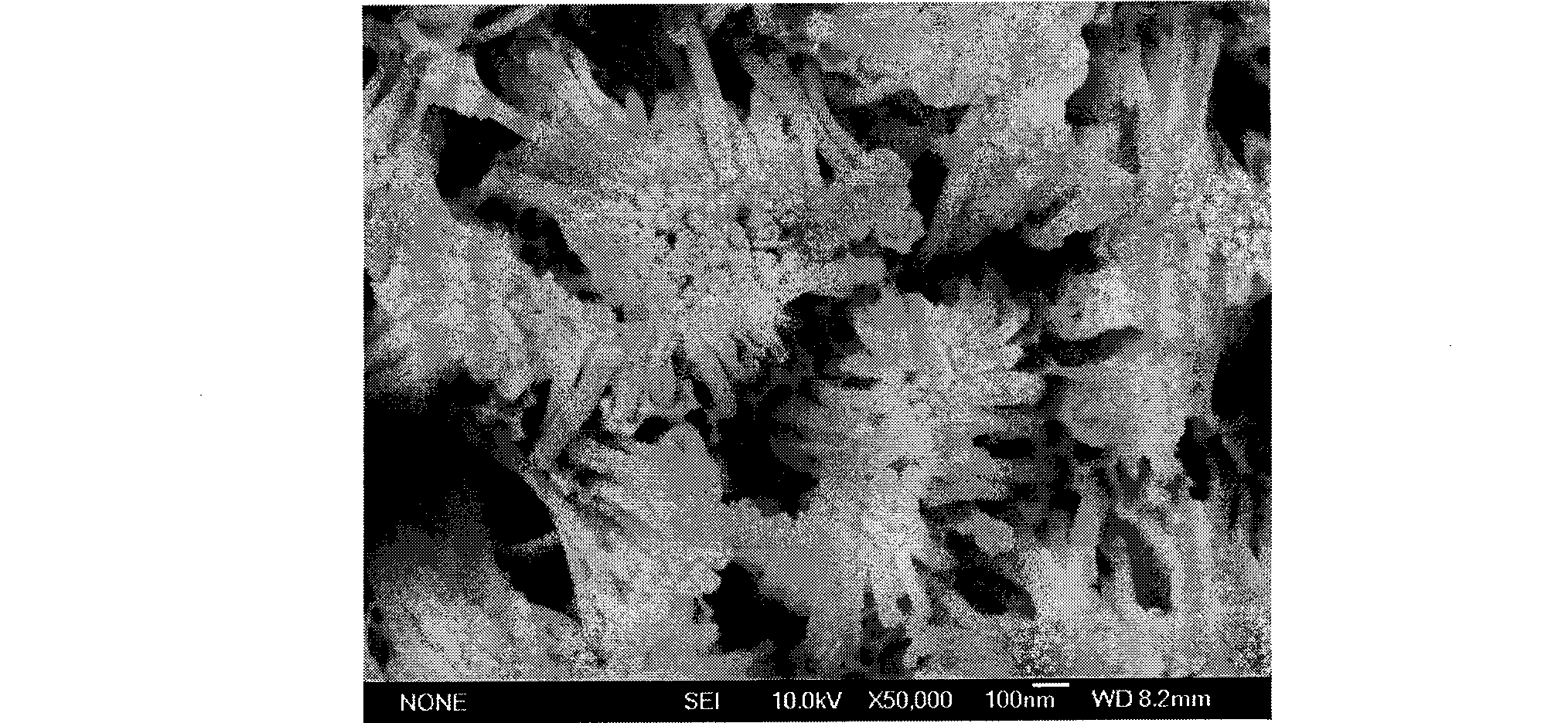
[0016] Take a piece of alumina template assembled with Te nanowire array, place it at the bottom of the beaker, use a dropper to take 0.1mL NaOH solution with a concentration of 1M and drop it in the middle of the template, after 5 minutes, inject 100mL NaOH solution with a concentration of 0.0001M After soaking for 15 minutes, the sample was taken out, washed several times with a large amount of water, dried and observed under a scanning electron microscope to obtain a scanning electron microscope photo of Te nanowire arrays treated by the method of the present invention. It can be seen from the scanning electron microscope photo in Figure 1 that the surface of the sample is smooth and clear, the holes of the template can be clearly seen, and no other...

Embodiment 2

[0019] Example 2: Ag in alumina template 2 Fabrication of SEM samples of Te nanowire arrays

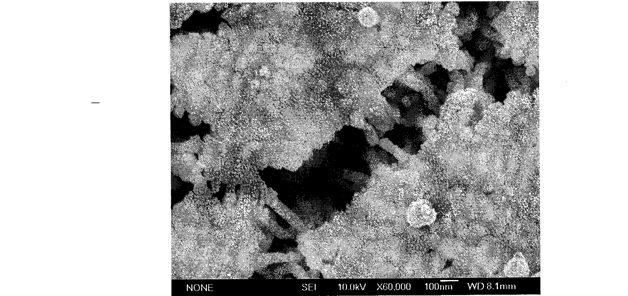
[0020] Take a piece assembled with Ag 2 Alumina template of Te nanowire array, place it at the bottom of the beaker, use a dropper to take 0.1mL of KOH solution with a concentration of 0.05M and drop it in the middle of the template, after 15 minutes, inject 20mL of KOH solution with a concentration of 0.0005M, soak for 15 After 10 minutes, take out the sample, wash repeatedly several times with a large amount of water, observe under the scanning electron microscope after drying, obtain the Ag after the inventive method process. 2 The scanning electron micrograph of Te nanowire array is shown in Fig. 3. It can be seen from the scanning electron microscope photo in Figure 3 that the surface of the sample is smooth and clear, and the details of the topography of the sample surface are not covered, and no other substances are found attached.

Embodiment 3

[0021] Example 3: Ag in alumina template 7 Te 4 Fabrication of SEM samples of nanowire arrays

[0022] Take a piece assembled with Ag 7 Te 4 The aluminum oxide template of the nanowire array is placed at the bottom of the beaker, and 0.05mL of NaOH solution with a concentration of 5M is dropped in the middle of the template with a dropper. After 30 minutes, 100mL of KOH solution with a concentration of 0.0001M is injected and soaked for 30 minutes. , take out the sample, wash it several times with a large amount of water, dry it and observe it under a scanning electron microscope, and get Ag 7 Te 4 The scanning electron micrograph of the nanowire array is shown in Fig. 4. It can be seen from the scanning electron microscope photo in Figure 4 that the surface of the sample is smooth and clear, and the details of the morphology are not covered up, and no other substances are found attached.

[0023] in conclusion:

[0024] As can be seen from the above examples, the scann...


PUM


Abstract

The invention discloses a method for manufacturing a scanning electron microscope sample for an assembled nanometer line array in an aluminum oxide template, which is characterized by comprising the following steps: eroding the template by an alkali liquor the concentration of which is between 0.05 and 5 M, soaking the template continuously in the alkali liquor with lower concentration of between0.001 and 0.0001 M for three to five minutes, then washing the template by water for a few times, and airing the template for the observation under the scanning electron microscope, wherein the alkali liquor is NaOH or KOH solution. The scanning electron microscope sample for the assembled nanometer line array in the aluminum oxide template manufactured by the method can avoid the effect caused by Al(OH)3 deposition attached on the surface of the sample on the observation of the scanning electron microscope.

Description

technical field [0001] The invention belongs to the technical field of manufacturing scanning electron microscope samples, and in particular relates to a manufacturing method of scanning electron microscope samples assembled with nanowire arrays in alumina templates. Background technique [0002] According to the U.S. "Physical Chemistry Journal B" (J.Phys.Chem.B, 2004, the 106th volume, the 1844th page) and the Dutch "Crystal Growth Journal" (J.Cryst.Growth, 2005, the 285th volume, the pp. 527 pages) report, the nanowire array assembled in the existing aluminum oxide template, the making of its scanning electron microscope sample all is to adopt lye (NaOH solution) to corrode the template, wash directly with water several times, and promptly put it under the scanning electron microscope after drying. observe. Since alumina reacts with alkali to produce metaaluminate, metaaluminate is easily hydrolyzed to generate Al(OH) when directly washed with water 3 Precipitation, the...

Claims


Application Information

Patent Timeline
Patent Type & Authority Patents(China)
IPC IPC(8): G01N1/32G01N13/10H01J37/28
Inventor 贾冲陈翌庆苏勇
Owner HEFEI UNIV OF TECH
Why Eureka
Try Eureka