
Bioabsorbable medical device with coating

A medical device, bioabsorption technology, applied in coating, medical science, surgery, etc.

Inactive Publication Date: 2009-10-21
ORBUSNEICH MEDICAL INC
View PDF6 Cites 6 Cited by
  • Summary
  • Abstract
  • Description
  • Claims
  • Application Information

AI Technical Summary

Problems solved by technology

One limitation of drug-eluting stents is that patients need to take supplemental oral medications such as aspirin and clopidogrel (clopidrogel) to prevent blood clots from developing early after implantation

Method used


Image

  • Bioabsorbable medical device with coating
  • Bioabsorbable medical device with coating
  • Bioabsorbable medical device with coating

Examples


Embodiment 1

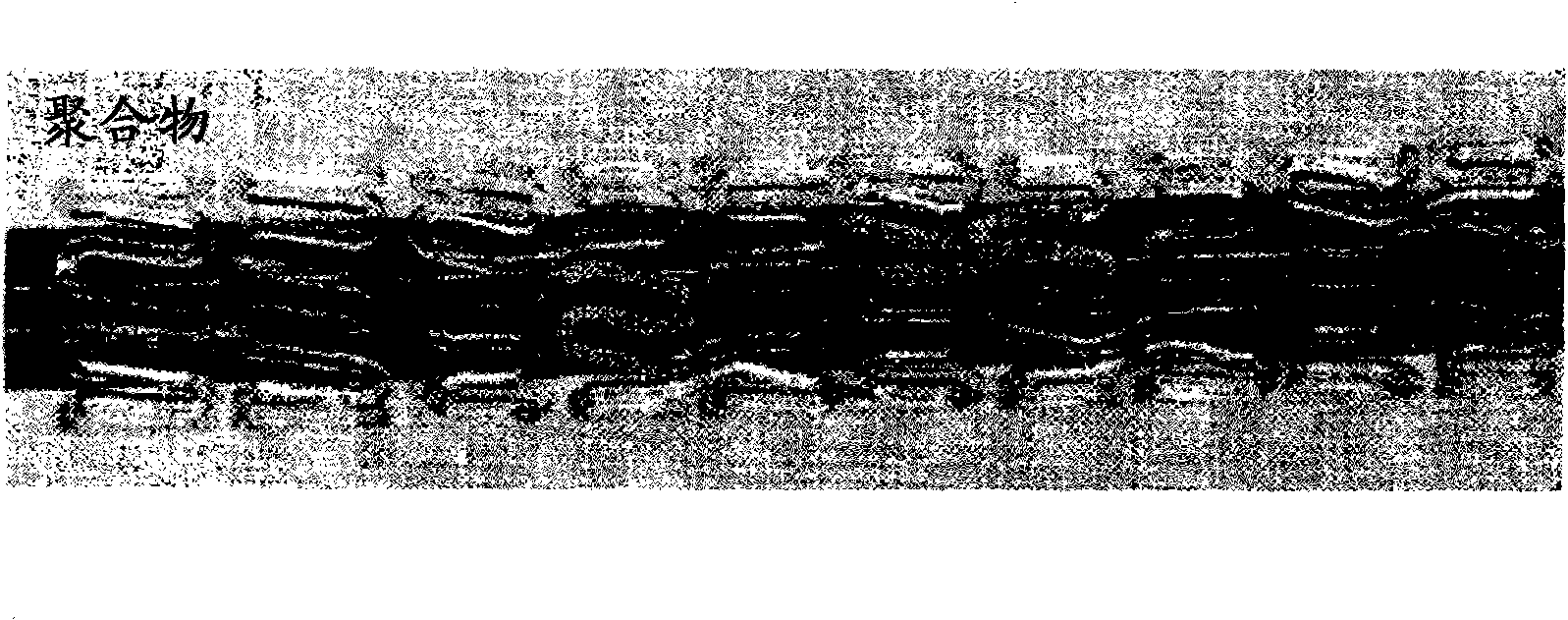
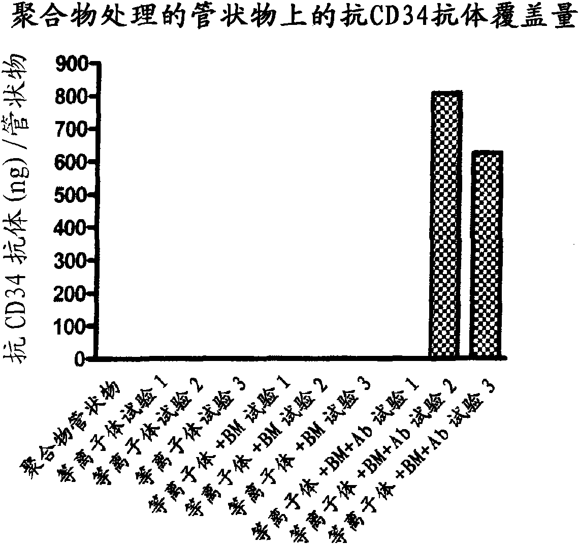
[0131] Bioabsorbable polymeric tubing used in the preparation of embodiments of the present invention is prepared from the polymer compositions described above. Uncoated tubing as well as tubing that had been coated as described above (comprising a coating with anti-CD34 antibody coated on a polymer matrix) were analyzed for their ability to bind antibodies to their surface. Prior to testing the tubing for cell binding, the antibody-coated tubing was checked for the amount of antibody bound to its surface. The experiment was repeated 3 times. The results of these studies are shown in figure 2 middle.

[0132] Such as figure 2 As shown in , the untreated tubing, the plasma treated (oxygen plasma followed by argon plasma) tubing, and the matrix-coated tubing did not each fail in any of the experiments performed. Contains any anti-CD34 antibody. In contrast, polymer tubes coated first with a coating solution comprising a bioabsorbable polymer and then with a buffer solutio...

Embodiment 2

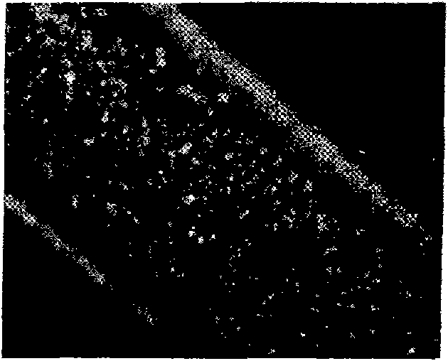
[0134] The non-coated and coated tubing as well as the plasma-treated tubing were incubated with Kg1a cells. Following incubation, the tubing was rinsed in buffered saline to remove unbound cells, and the tubing was fixed and processed to identify cells bound to the device. Tested tubules were stained by fluorescent (nucleostain DAPI ((4',6-diamidino-2-phenylindole) dihydrochloride)) staining after incubation and examined under a fluorescent microscope Binding of cells to the tubing was determined. The results of these experiments are shown in Figure 3-6 .

[0135] image 3 A representative bioabsorbable tube is shown with a coating comprising polymer and anti-CD34 antibody, depicting the adhesion of many Kg1a cells to the tube, which can be visualized by the fluorescence emitted from the cells. Figure 4 is a representative non-coated tubing from the experiment showing few adherent cells on the non-coated tubing. Most of the signal from these panels appeared to be due t...


PUM


Abstract

A biodegradable, bioabsorbable medical device with a coating for capturing progenitor endothelial cells in vivo and delivering a therapeutic agent at the site of implantation. The coating on the medical device is provided with a bioabsorbable polymer composition such as a bioabsorbable polymer, copolymer, or terpolymer, and a copolymer or terpolymer additive for controlling the rate of delivery ofthe therapeutic agent.

Description

[0001] Cross references to related patent applications [0002] This application claims priority based on US Provisional Patent Application Serial No. 60 / 862,409 filed October 20,2006. Background technique [0003] All references cited in this specification and their references are hereby incorporated by reference in their entirety where appropriate for teaching additional or alternative details, features and / or technical background. [0004] In the embodiments disclosed herein, the invention relates to a novel medical device having a coating. Such devices may be shaped for implantation into vascular or luminal structures in the body. More specifically, in embodiments, the present invention relates to stents and synthetic grafts coated with a controlled release matrix comprising a pharmaceutical substance for direct delivery to surrounding tissue, and attached thereto Ligands for capturing endothelial progenitor cells that may be present in bodily fluids that contact the sub...

Claims


Application Information

Patent Timeline
Patent Type & Authority Applications(China)
IPC IPC(8): A61L27/28
CPCA61L27/34A61L2300/80A61L31/06A61L27/18A61L31/16A61L27/58A61L31/18A61L27/54A61L31/10A61L27/507A61L31/148
Inventor R·J·卡特恩
Owner ORBUSNEICH MEDICAL INC
Learn More