Method for marking bifluorescence protein molecule cell

A labeling method and dual-fluorescence technology, which can be applied to cells modified by introducing foreign genetic material, introducing foreign genetic material using a vector, recombinant DNA technology, etc. It can solve the problems of labeling that are not suitable for living cells, and save the time of labeling. , the effect of simple marking process

Inactive Publication Date: 2009-11-04
SHANGHAI INST OF BIOLOGICAL SCI CHINESE ACAD OF SCI
View PDF0 Cites 8 Cited by
  • Summary
  • Abstract
  • Description
  • Claims
  • Application Information

AI Technical Summary

Problems solved by technology

[0003] However, because the antibody method cannot freely enter or cells, it is not suitable for the labeling of living cells, and the GFP method can be used for the labeling of living cells, but because the GFP-fused ...

Method used


Image

  • Method for marking bifluorescence protein molecule cell
  • Method for marking bifluorescence protein molecule cell
  • Method for marking bifluorescence protein molecule cell

Examples


Embodiment 1

[0019] Example 1 , Construction of recombinant plasmids

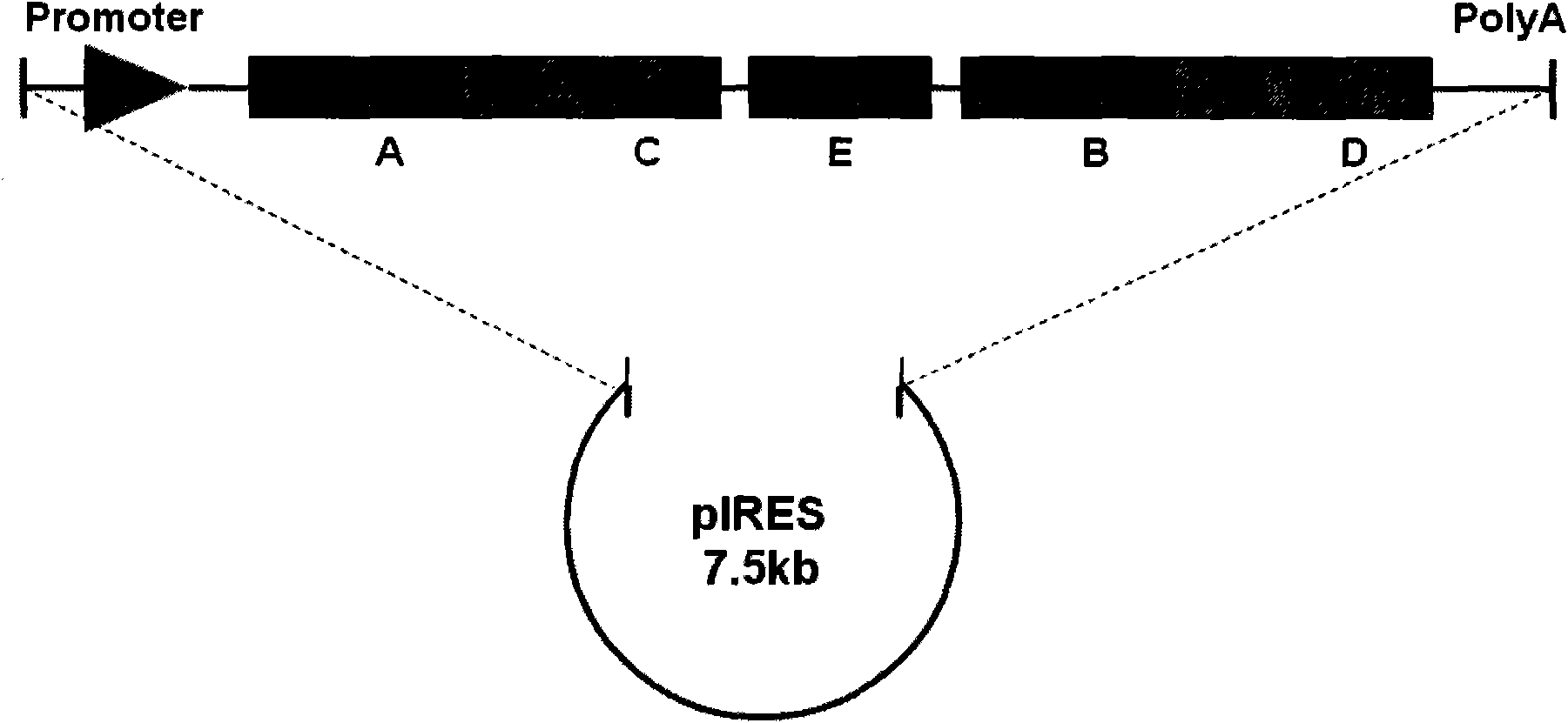
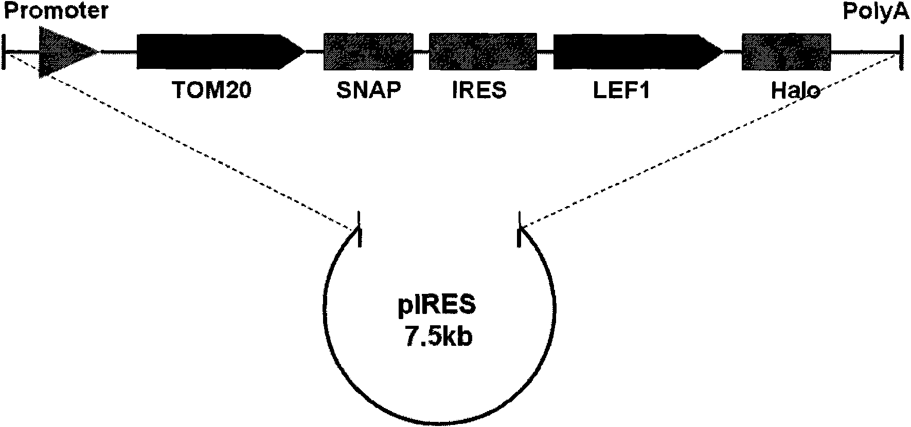
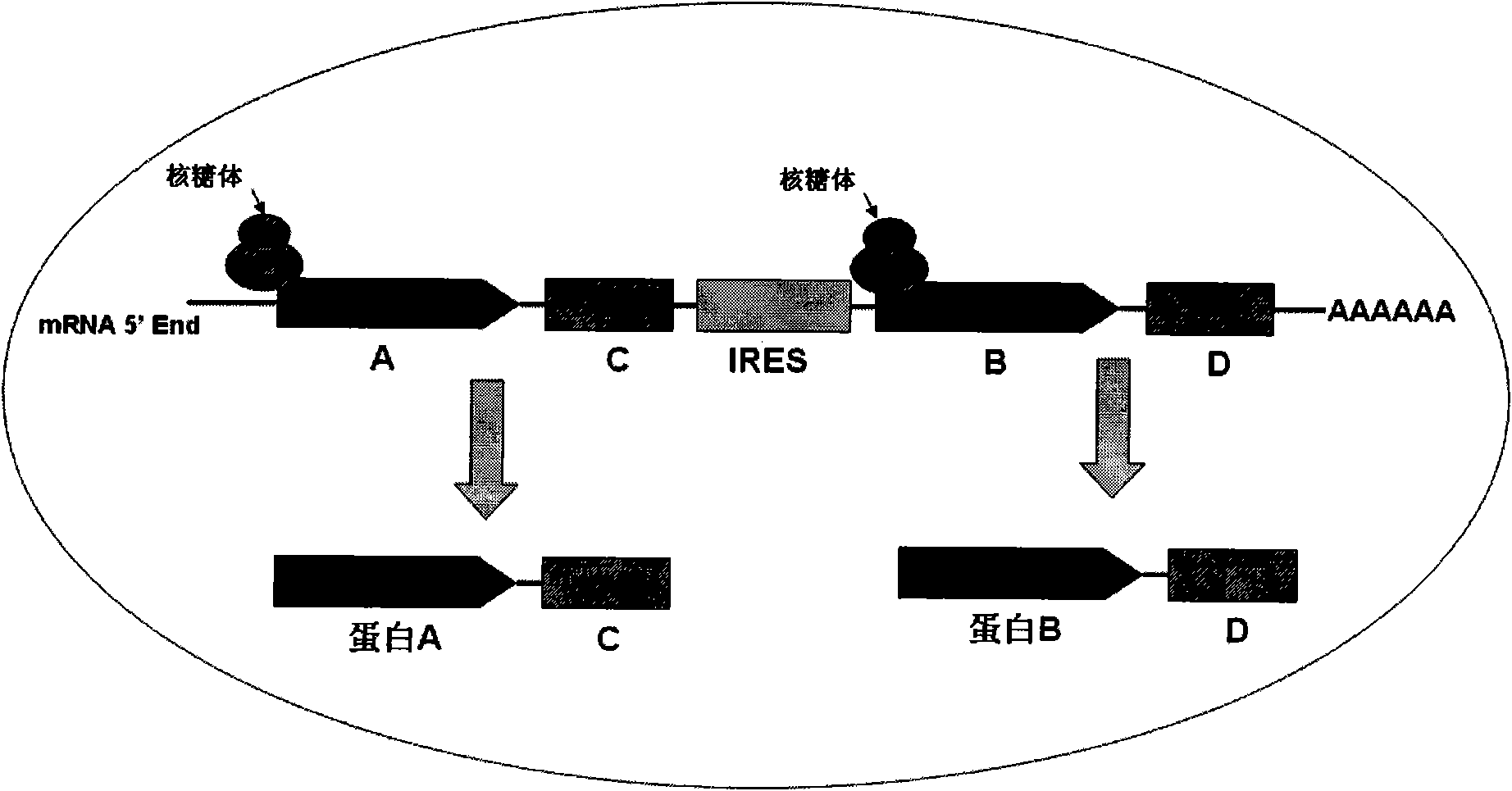
[0020] In this example, the inventor constructed the pTOM20-SNAP-IRES-LEF1-Halo recombinant plasmid, in which the inventor cloned the pTOM20-SNAP-IRES-LEF1-Halo fragment, wherein the protein LEF1 is the Wnt signal The protein molecules in the pathway are mainly located in the nucleus. The protein TOM20 is a protein on the outer membrane of the mitochondria, which is located on the mitochondria; IRES is a bicistronic ORF expression system of the internal ribosome insertion site element, making the two genes Two ORFs are co-expressed under the same promoter in one vector, the first cistron expression cassette is fused with SNAP at the C-terminus, and the second is fused to express Halo.

[0021] The construction of the pTOM20-SNAP-IRES-LEF1-Halo fragment includes the following 4 steps: 1. Connect the LEF1 obtained by PCR into the pFC14A vector with Halo to obtain the pFC14A-LEF1 recombinant plasmid; 2. Digest the LEF1- ...

Embodiment 2

[0052] Example 2 , transfected cells and fluorescent labeling

[0053] 2.1. Cell transfection

[0054] Use the Lipo-plus method to transfect Hela cells, the specific steps are as follows:

[0055] (1) Put the slides cleaned and sterilized with medical alcohol into a 24-well plate, coat them with poly-L-Lysine (Poly-L-Lysine) for 30 minutes, wash them with sterilized water for 3-5 times, and wash away Uncoated PLL, dry on a clean bench;

[0056] (2) Hela cells were cultured for 24 hours in a 24-well plate coated with PLL slides;

[0057] (3) Take 250ng of pTOM20-SNAP-IRES-LEF1-Halo recombinant plasmid and add it to the EP tube containing 25ul serum-free DMEM culture medium, and mix well;

[0058] (4) Take the transfection reagent plus, dilute it into DMEM culture medium at 1:20, mix well, add 21ul / tube into the EP tube in the previous step, mix well, and let stand for 15 minutes;

[0059] (5) Take the transfection reagent lipo, dilute it into DMEM culture medium at 1:20, ...


PUM


Abstract

The invention provides a recombinant plasmid for a method for marking a bifluorescence protein molecule and a method for marking a bifluorescence protein molecule cell. The recombinant plasmid comprises a segment A coding tag protein A, a segment B coding tag protein B and a promoter at the upper stream of a segment AC, wherein the 5' end or the 3' end of the segment A merges a sequence C coding protein C to be detected; the 5' end or the 3' end of the segment B merges a sequence D coding protein D to be detected; and segments A-C and B-D are connected by an IRES sequence E. The marking method comprises the steps of constructing the recombinant plasmid for the method for marking the bifluorescence protein molecule, cell transfection and fluorescence marking. The recombinant plasmid and the marking method can express two target genes in all in the same carrier, marks two kinds of different proteins in the same cell, has simple and rapid marking process and greatly saves the marking time than that of a traditional antibody fluorescence marking method.

Description

technical field [0001] The invention belongs to the field of biotechnology, and in particular relates to a method for cell labeling with dual fluorescent protein molecules. Background technique [0002] Fluorescence microscopy provides an extremely useful tool for the study of cell biology. Through intuitive images, the dynamic behavior of protein molecules in cells can be studied. Such as protein secretion, endocytosis, transport, localization, protein export and import into nucleus, interaction of protein molecules in cells, etc. The traditional method is through the fusion expression of protein molecules and tag proteins (such as GFP, YFP, Flag, HA, etc.), in cells through the autofluorescence of GFP or the fluorescent molecules coupled with the corresponding antibodies of various tag proteins Fluorescence of various wavelengths emitted after being excited achieves the purpose of observation. [0003] However, because the antibody method cannot freely enter or cells, it...

Claims


Application Information

Patent Timeline
IPC IPC(8): C12N15/63C12N15/65C12N5/10
Inventor 杨淑伟黄文韬
Owner SHANGHAI INST OF BIOLOGICAL SCI CHINESE ACAD OF SCI
Why Eureka
Try Eureka