Integrated SPECT imaging and ultrasound therapy system

An imaging subsystem and ultrasound technology, applied in ultrasound therapy, treatment, ultrasound/sonic/infrasonic diagnosis, etc., can solve problems such as coordination difficulties

Inactive Publication Date: 2009-11-04
KONINKLIJKE PHILIPS ELECTRONICS NV
View PDF0 Cites 8 Cited by
  • Summary
  • Abstract
  • Description
  • Claims
  • Application Information

AI Technical Summary

Problems solved by technology

However, imaging techniques that will provide accurate images in a well-defined coordinate system, such as CT scans, can be difficult to coordinate with manual treatment delivery systems

Method used


Image

  • Integrated SPECT imaging and ultrasound therapy system
  • Integrated SPECT imaging and ultrasound therapy system
  • Integrated SPECT imaging and ultrasound therapy system

Examples


Embodiment Construction

[0015] The present disclosure relates to advantageous integration and co-registration of ultrasound transducers with SPECT imaging systems. The disclosed SPECT imaging system can be used with a wide range of contrast agents to provide highly sensitive and specific detection of disease. The disclosed ultrasound transducers allow delivery of particle-born or microbubble-based drug therapy or thermoactive therapy at the same sites detected with SPECT imaging. The integration of these two modalities can be used to substantially improve patient care through, inter alia, improved spatial alignment, treatment planning, and workflow.

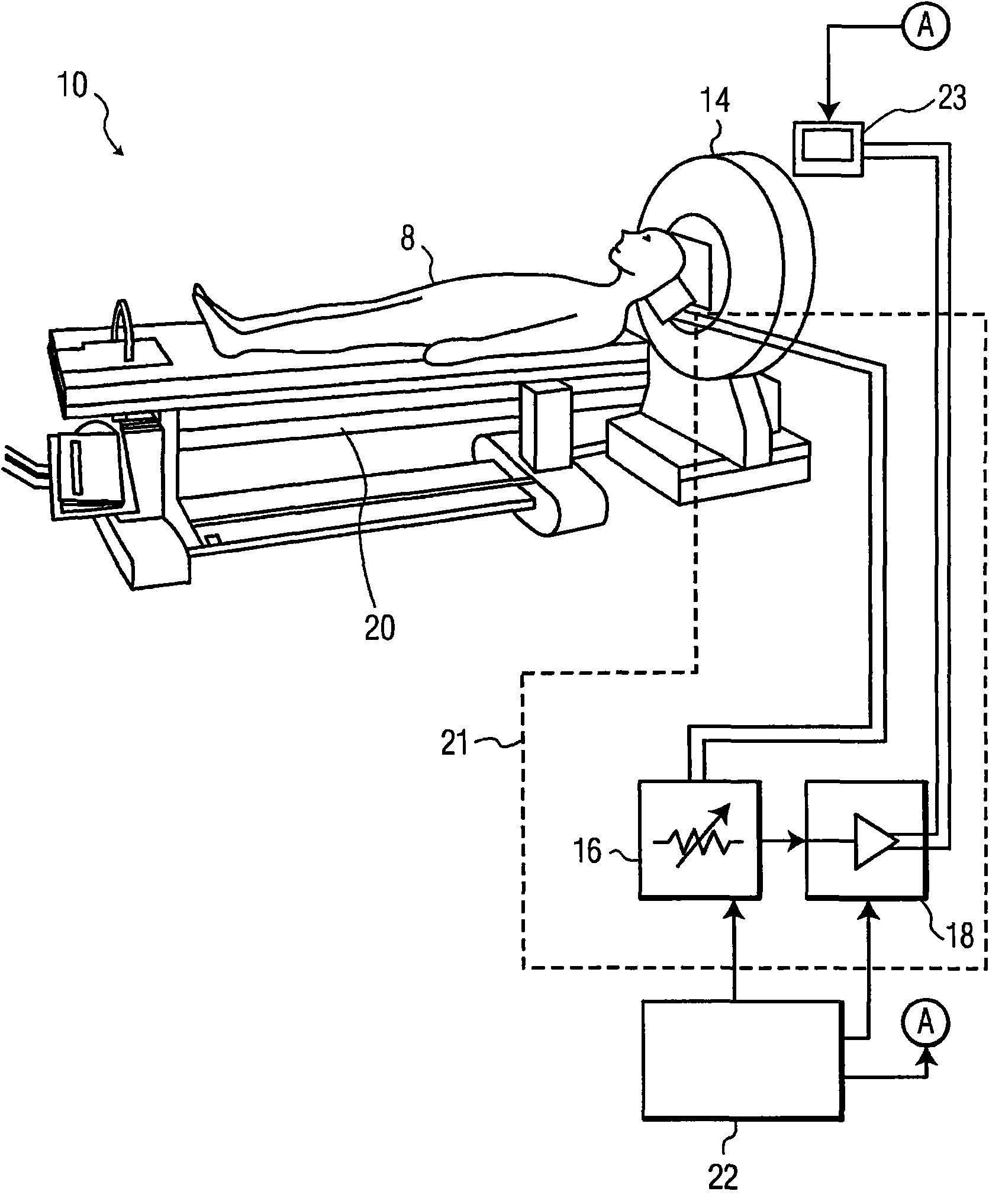
[0016] refer to figure 1 , shows a schematic diagram of an exemplary system integrating a SPECT imaging instrument and an ultrasound transducer, generally indicated at 10 , according to the present disclosure. The system 10 includes an ultrasound transducer 12, a SPECT imaging subsystem 14 for controlling an ultrasound beam generated by the transducer...


PUM


Abstract

A method and apparatus for the integration of an ultrasound transducer with a SPECT imaging system is disclosed. The ultrasound transducer allows the delivery of particle-born drug therapies or thermally active therapies at the same site detected with SPECT imaging. The system includes a SPECT imaging subsystem; an ultrasound transducer for producing a signal representative of applied ultrasound and signally coupled to the SPECT imaging subsystem; and co-registering means for co-registering the signal representative of applied ultrasound in a coordinate system of SPECT imaging subsystem. The SPECT subsystem may be replaced with a SPECT / CT combined system to provide morphology that allows for treatment planning. In another embodiment, the ultrasound transducer can be used intermittently with the sequential gathering of SPECT images for tracking time-varying properties of the binding of specific SPECT contrast agents to biomarkers of disease. In this way, the ultrasound and SPECT imaging devices form a novel combination to improve patient care through improved spatial registration, therapy planning, and workflow.

Description

technical field [0001] The present disclosure relates to an apparatus and method for integrating single photon emission computed tomography (SPECT) with an ultrasound transducer. Background technique [0002] Molecular imaging technologies are revolutionizing the clinical approach to diagnosis and staging of specific pathologies. In these techniques, imaging modality-specific contrast agents are often combined with supplements for biomarkers of disease. These markers are usually proteins expressed during various stages of the disease, but sometimes they can also be metabolites of altered cells associated with pathological cells. Contrast agents are specific to each modality. One imaging modality that holds great promise for the future of molecular imaging is single photon emission computed tomography (SPECT). SPECT also has the ability to model the flow of fluids in three dimensions, which enables it to image blood flow and the delivery of therapeutic drugs to target loca...

Claims


Application Information

Patent Timeline
Patent Type & Authority Applications(China)
IPC IPC(8): A61B8/00
CPCA61N7/02A61B6/037
Inventor C·S·霍尔S·索卡B·I·拉朱
Owner KONINKLIJKE PHILIPS ELECTRONICS NV
Why Eureka
Try Eureka