DDK-1 specific siRNA and application thereof in treatment of rheumatoid arthritis
A DKK-1, rheumatoid technology, applied in the direction of DNA / RNA fragments, recombinant DNA technology, gene therapy, etc., can solve the problems of restricting the development of treatment methods and unclear pathogenesis
- Summary
- Abstract
- Description
- Claims
- Application Information
AI Technical Summary
Problems solved by technology
Method used
Image
Examples
Embodiment 1
[0015] Example 1: Screening and identification of siRNA
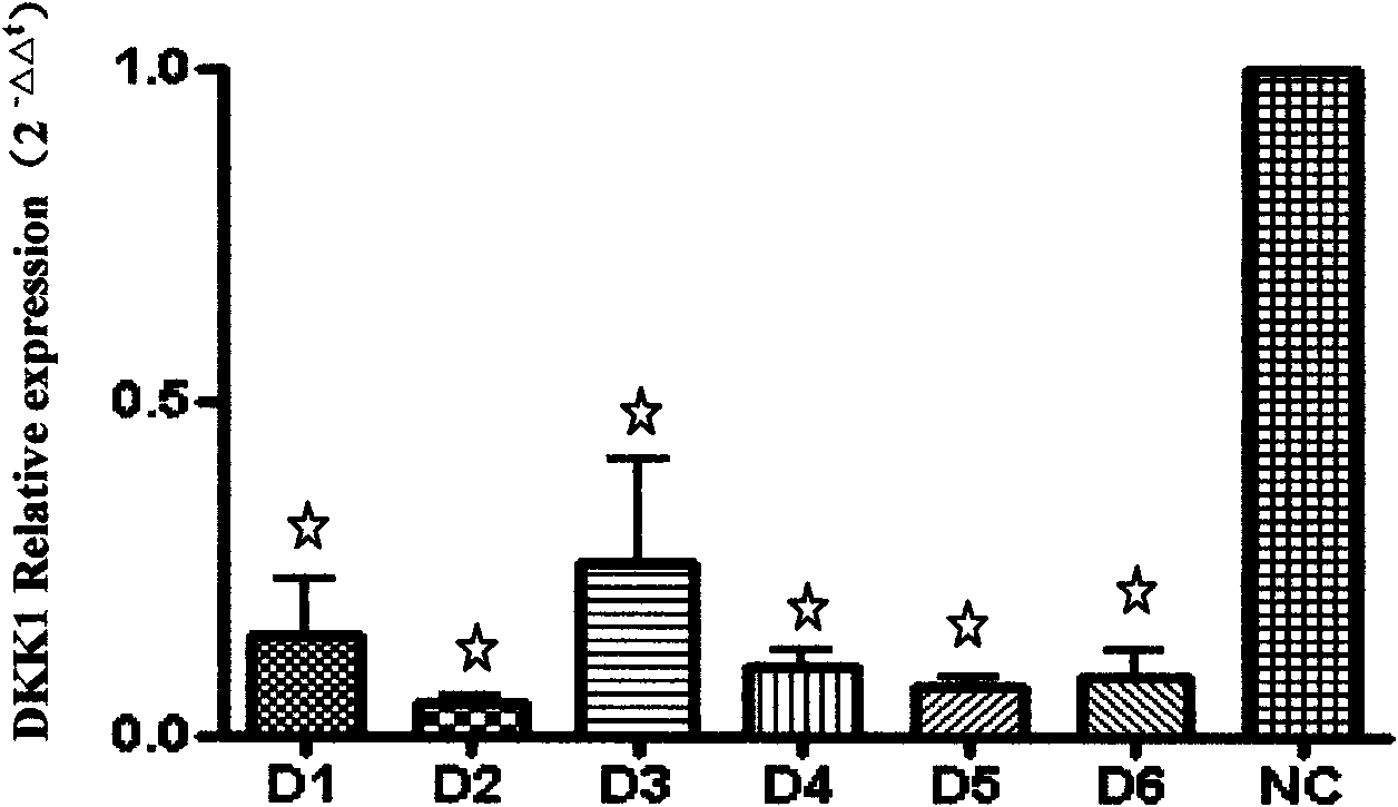
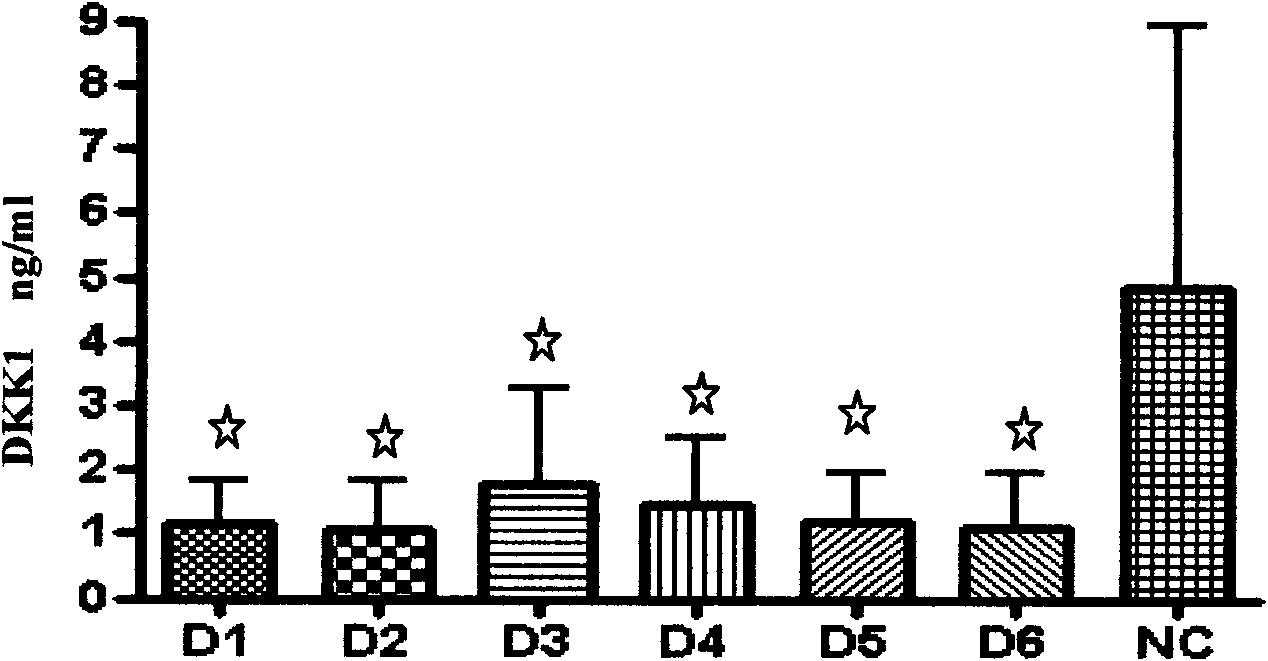
[0016] Collect the synovial tissue after knee joint replacement in RA patients, isolate and culture synovial fibroblasts, and use the cells after 3 generations for experiment. Spread in a 12-well plate, 5×10 cells per well 4 One, set up multiple holes. After 48 hours, the cells were collected, the total RNA was extracted by Trizol method, and A260 was measured with an ultraviolet spectrophotometer. The relative expression level of DKK-1 uses ΔCt and 2 -ΔΔCt For calculation, GAPD is used as an internal reference. Real-time quantitative PCR uses SYBR from ABI Green PCR Master Mix is operated. ELISA method was used to detect the expression level of DKK-1 in the cell supernatant. The results showed that after the 6 pairs of siRNAs designed were transferred into the cells, they could significantly inhibit the expression of DKK-1, and a pair of siRNAs with high inhibition efficiency was screened, as shown in Figure 1 and ...
Embodiment 2
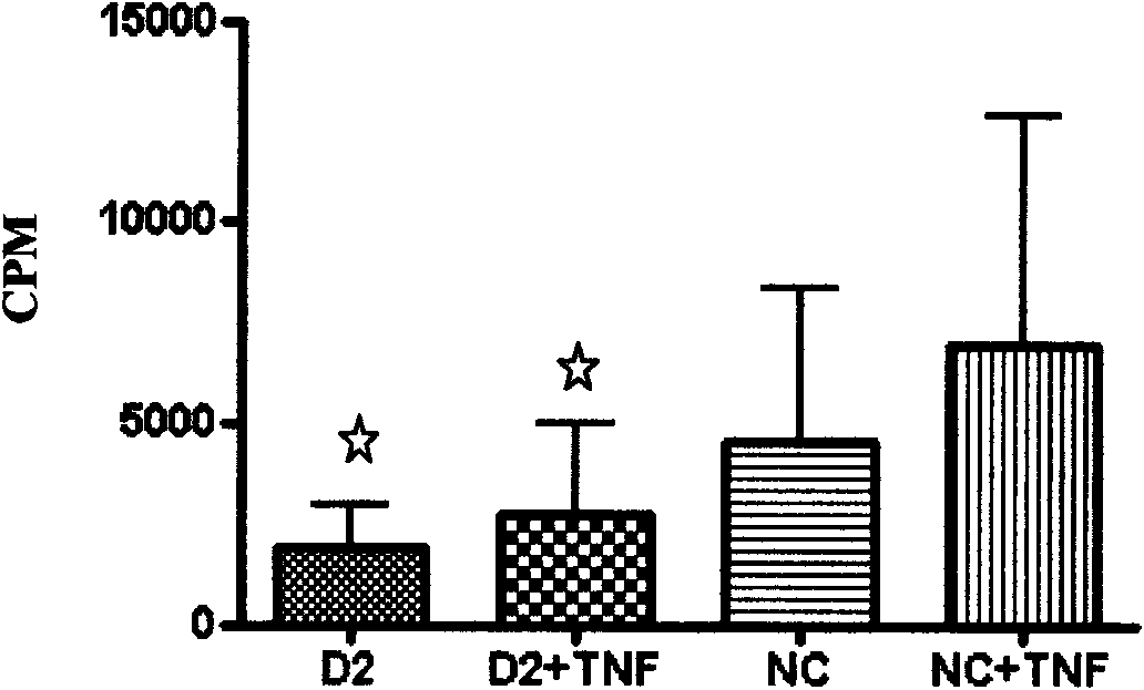
[0017] Example 2: The effect of siRNA targeting DKK-1 on the proliferation ability of RA-FLS
[0018] Isolate and culture synovial fibroblasts and divide them into two groups, one group is no TNF-α activation group, and the other is TNF-α activation group (10ng / ml). Lipo2000 liposome transfection and chemical synthesis of siRNA were performed at the same time as control. Use unrelated sequence siRNA. Use 96-well plates for transfection, 5×10 cells per well 3 , Set up a complex hole. Collect the cells after 48 hours, 3 The H incorporation method was used to detect the proliferation of fibroblast-like synovial cells. The results showed that the proliferation of FLS in the TNF-α activated group was more obvious, but after transfection of siRNA, the proliferation of FLS in the two groups was significantly inhibited, as shown in Figure 3.
Embodiment 3
[0019] Example 3: The effect of down-regulating the expression of DKK-1 on the secretion of inflammatory factors by RA-FLS
[0020] Use FLS of RA patients after 3 generations, 5×10 4 / Well was inoculated into a 12-well plate, Lipo2000 liposomes were transfected with chemically synthesized siRNA and negative control, and then divided into TNF-α induction (10ng / ml) and no TNF-α groups. Each group was a duplicate well and was detected by ELISA The secretion levels of IL-6, IL-8, MMP2 and MMP9 in FLS supernatant 48h after transfection. The results showed that FLS secreted more inflammatory factors in the TNF-α activated group, but after transfection of siRNA, the secretion of FLS inflammatory factors in the two groups had a significant inhibitory effect. Figure 4, Figure 5, Figure 6 and Figure 7.
PUM
 Abstract
Description
Claims
Application Information
 - R&D
- Intellectual Property
- Life Sciences
- Materials
- Tech Scout
- Unparalleled Data Quality
- Higher Quality Content
- 60% Fewer Hallucinations
Browse by: Latest US Patents, China's latest patents, Technical Efficacy Thesaurus, Application Domain, Technology Topic, Popular Technical Reports.
© 2025 PatSnap. All rights reserved.Legal|Privacy policy|Modern Slavery Act Transparency Statement|Sitemap|About US| Contact US: help@patsnap.com


