Retinal image segmentation method based on NSCT feature extraction and supervised classification

A technology of feature extraction and supervised classification, applied in the field of image processing, can solve problems such as error, poor contrast between blood vessels and background, and achieve good segmentation accuracy and small error

Inactive Publication Date: 2010-03-10
XIDIAN UNIV
View PDF0 Cites 19 Cited by
  • Summary
  • Abstract
  • Description
  • Claims
  • Application Information

AI Technical Summary

Problems solved by technology

[0006] In retinal image blood vessel segmentation, the most important thing is to increase the detection rate of blood vessels. However, due to the global unbalanced gray scale of retinal images, poor contrast between blood ve

Method used


Image

  • Retinal image segmentation method based on NSCT feature extraction and supervised classification
  • Retinal image segmentation method based on NSCT feature extraction and supervised classification
  • Retinal image segmentation method based on NSCT feature extraction and supervised classification

Examples


Embodiment Construction

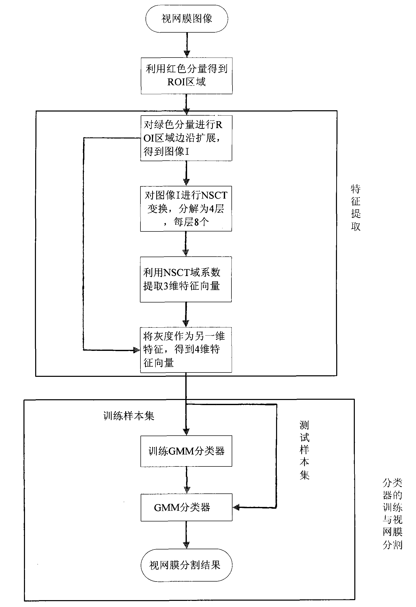
[0031] refer to figure 1 , the specific implementation process of the present invention is as follows:

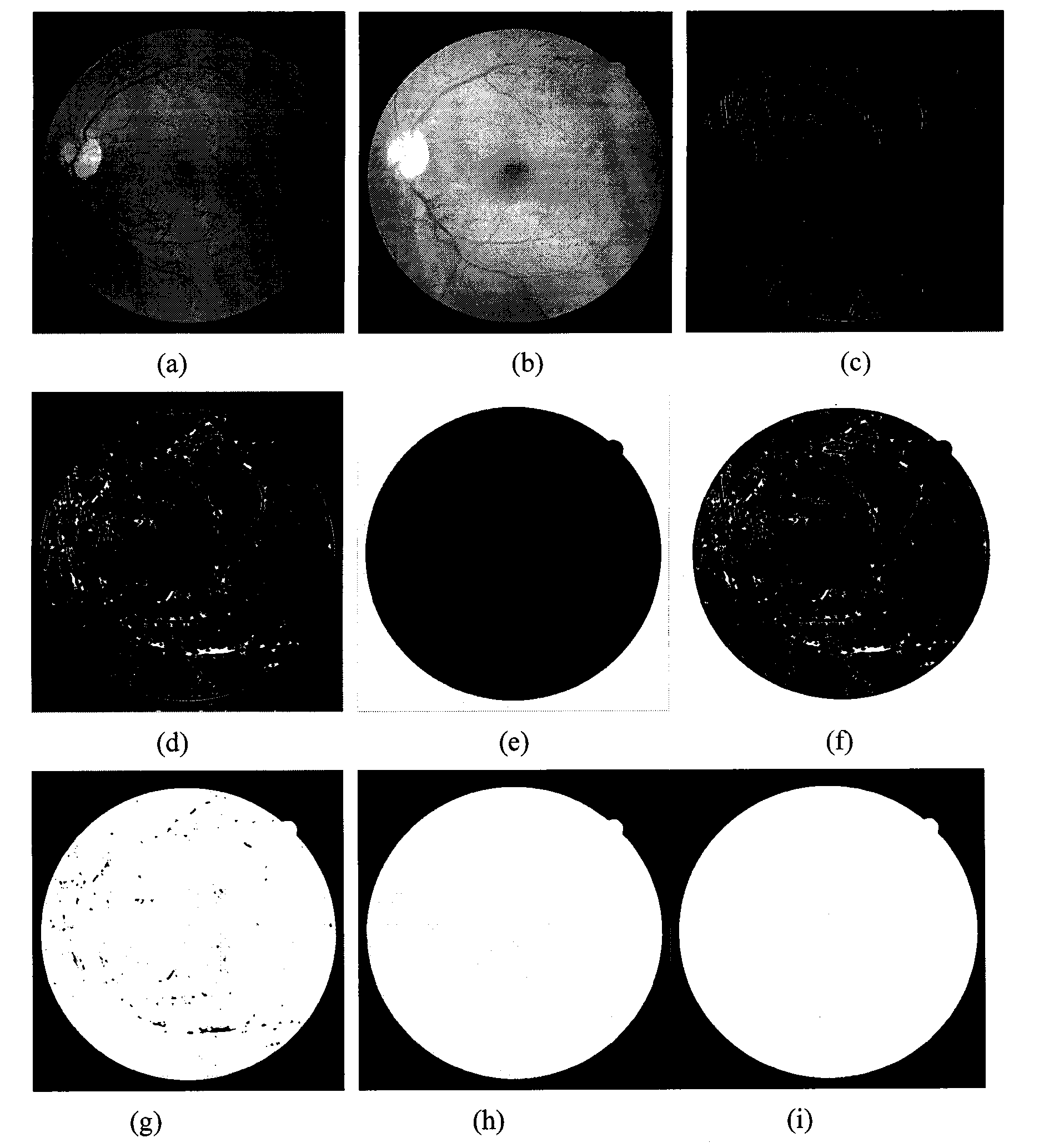
[0032] Step 1: Use the red component of the retinal training image and the retinal image to be segmented to obtain the region of interest ROI. For specific steps, refer to figure 2 as follows:

[0033] (1.1) For example figure 2 The red component of the retinal image shown in (a) is divided by 255, as figure 2 (b), and through the Gaussian filter LOG pair figure 2 (b) Perform edge detection to get figure 2 (c);

[0034] (1.2) Yes figure 2 (c) Carry out first expansion and then corrosion, so that the edge fractures are connected to obtain figure 2 (d);

[0035] (1.3) in figure 2 In (d), add a contour along the edge of the image;

[0036] (1.4) Determine the outer area by threshold, in figure 2 In (b), find the maximum value of its grayscale max red, and mark the points whose grayscale is smaller than max red×0.15 as 1, thus obtaining a binary image, and t...


PUM


Abstract

The invention discloses a retinal image segmentation method based on NSCT feature extraction and supervised classification, which relates to medical image processing. The method comprises the following steps: (1) obtaining interest regions of a retinal training image and a retinal image to be segmented by utilizing red components of the retinal training image and the retinal image to be segmented;(2) respectively carrying out edge iterative expansion of the regions of interest on green components of the retinal training image and the retinal image to be segmented; (3) respectively carrying out NSCT transformation on the expanded images to decompose the images into i layers; (4) extracting a one-dimensional features by utilizing sub-band coefficients in j directions of each layer, extracting the features layer by layer to form feature vectors and normalizing the feature vectors; (5) establishing a training sample of the feature vectors of the normalized retinal training image; (6) selecting a classifier, training the classifier by utilizing the training sample and inputting the feature vectors of the normalized retinal image to be segmented into the classifier so as to segment theretinal image to be segmented. The invention has the advantages of clear image segmentation edge and high precision and is used for medical image retina segmentation.

Description

technical field [0001] The invention belongs to the technical field of image processing, relates to the application of retinal detection, and can be used to extract retinal blood vessels from retinal images in medicine. Background technique [0002] The development of medicine is closely related to human health, so digital image processing technology has aroused great interest in biomedical circles from the very beginning. As early as the end of the 1970s, literature statistics pointed out that a very wide application of image processing is medical image processing. In medicine, whether it is in basic disciplines or clinical applications, it is a field with many types of image processing. However, due to the technical difficulty of medical image processing, it is difficult to achieve clinical practicality in many processes. In recent years, with the reduction of the cost of digital image processing equipment, the use of digital image processing technology to improve the qu...

Claims


Application Information

Patent Timeline
IPC IPC(8): G06T7/00G06K9/62A61B3/10
Inventor 钟桦焦李成侯鹏王爽侯彪刘芳马文萍公茂果
Owner XIDIAN UNIV
Why Eureka
Try Eureka