Laser imaging method and device for automatically cutting artery blood vessel and vein blood vessel

An automatic segmentation and laser imaging technology, applied in the field of biomedical imaging, can solve problems such as automatic separation of arterial blood vessels and venous blood vessels, and achieve the effect of wide application range and avoiding complexity.

Active Publication Date: 2010-04-28
HUAZHONG UNIV OF SCI & TECH
View PDF0 Cites 17 Cited by
  • Summary
  • Abstract
  • Description
  • Claims
  • Application Information

AI Technical Summary

Problems solved by technology

However, due to the complexity of the regional distribution of blood flow velocity, the complexity of the speckle contrast distributi

Method used


Image

  • Laser imaging method and device for automatically cutting artery blood vessel and vein blood vessel
  • Laser imaging method and device for automatically cutting artery blood vessel and vein blood vessel
  • Laser imaging method and device for automatically cutting artery blood vessel and vein blood vessel

Examples


Embodiment Construction

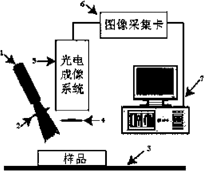
[0033] The laser imaging method and device for automatically segmenting the arteriovenous vessels of biological tissues proposed by the present invention adopt the following methods: figure 1 The imaging device shown has the following structure: the laser beam 1, the first linear polarizer 2 and the workbench 3 are sequentially located on the illumination optical path, and the first linear polarizer 2 is perpendicular to the incident laser beam 1; the workbench 3, the second The polarizer 4 and the photoelectric imaging system 5 are located on the imaging optical path in turn, the second linear polarizer 4 is perpendicular to the optical axis direction of the photoelectric imaging system 5, concentric with the photoelectric imaging system 5, and its polarization direction is the same as that of the first linear polarizer 2 Vertical; the computer 7 is connected to the photoelectric imaging system 5 through the image acquisition card 6, and the computer 7 controls the photoelectr...


PUM


Abstract

The invention discloses a laser imaging method and a device for automatically cutting artery blood vessel and vein blood vessel. The invention adopts single wavelength laser to illuminate; based on the difference of dynamic characteristics and spectral absorption characteristics of laser speckle of an artery zone and a vein zone in biological tissues, artery blood vessels and vein blood vessels are automatically cut by operations, such as time sequence image pixel gray degree minimum analysis, background non-uniformity correction and the like. The invention is suitable for physiology, pathology, pharmacology, drug effect evaluation research as well as clinical medical diagnosis and treatment.

Description

technical field [0001] The invention belongs to biomedical imaging technology method, specifically a method and device for automatic segmentation of arteriovenous blood vessels of biological tissue based on laser speckle image time series analysis using single-wavelength coherent light illumination, which is especially suitable for research with high spatial resolution Arterial and venous vessel morphology and blood flow distribution in biological tissue under physiological and pathological conditions provide important information for basic research in life sciences and clinical medical diagnosis and treatment. Background technique [0002] Automatic segmentation of arteriovenous vessels in biological tissues is of great significance for biomedical research and clinical diagnosis and treatment. Currently, there are very limited reports on methods for fully automatic segmentation of arteriovenous vessels. Arterial and venous blood vessels due to oxygenated hemoglobin (HbO 2...

Claims


Application Information

Patent Timeline
IPC IPC(8): A61B5/00G06T1/00G06T5/00
Inventor 李鹏程骆清铭邱建军
Owner HUAZHONG UNIV OF SCI & TECH
Why Eureka
Try Eureka