Integrated rigid ultrasonic cystoscope system

An ultrasound system and cystoscope technology, applied in the directions of ultrasound/sonic/infrasonic diagnosis, laparoscopy, endoscopy, etc., can solve the problems of affecting the quality of diagnosis, instability, inconvenient operation, etc., to improve image quality, not easy damage, and the effect of improving accuracy

Inactive Publication Date: 2010-08-18
广州市番禺区胆囊病研究所
View PDF6 Cites 3 Cited by
  • Summary
  • Abstract
  • Description
  • Claims
  • Application Information

AI Technical Summary

Problems solved by technology

However, the micro-ultrasound probe is easy to be damaged due to improper use during use, and because the micro-ultrasound probe is used in conjunction with the endoscope due to inconvenient operation and instability, it is easy to cause asynchron

Method used


Image

  • Integrated rigid ultrasonic cystoscope system
  • Integrated rigid ultrasonic cystoscope system
  • Integrated rigid ultrasonic cystoscope system

Examples


Embodiment Construction

[0019] Below in conjunction with accompanying drawing, the present invention is described in further detail:

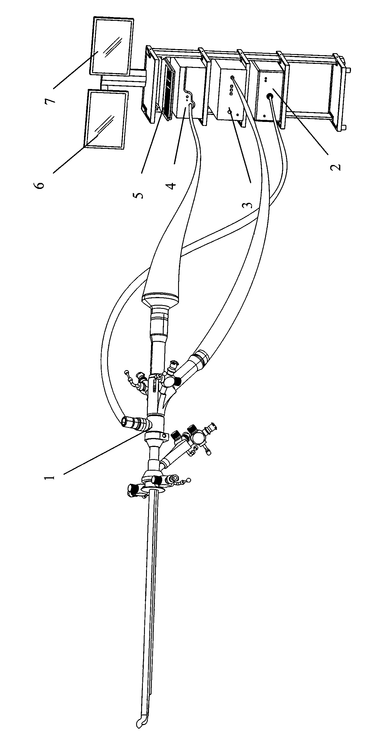
[0020] Such as figure 1 As shown, the integrated rigid ultrasonic cystoscope system of the present invention includes an integrated rigid ultrasonic cystoscope 1, a light source host 2, a camera host 4, an ultrasound system host 3 and a keyboard 5, two medical monitors 6 and 7 .

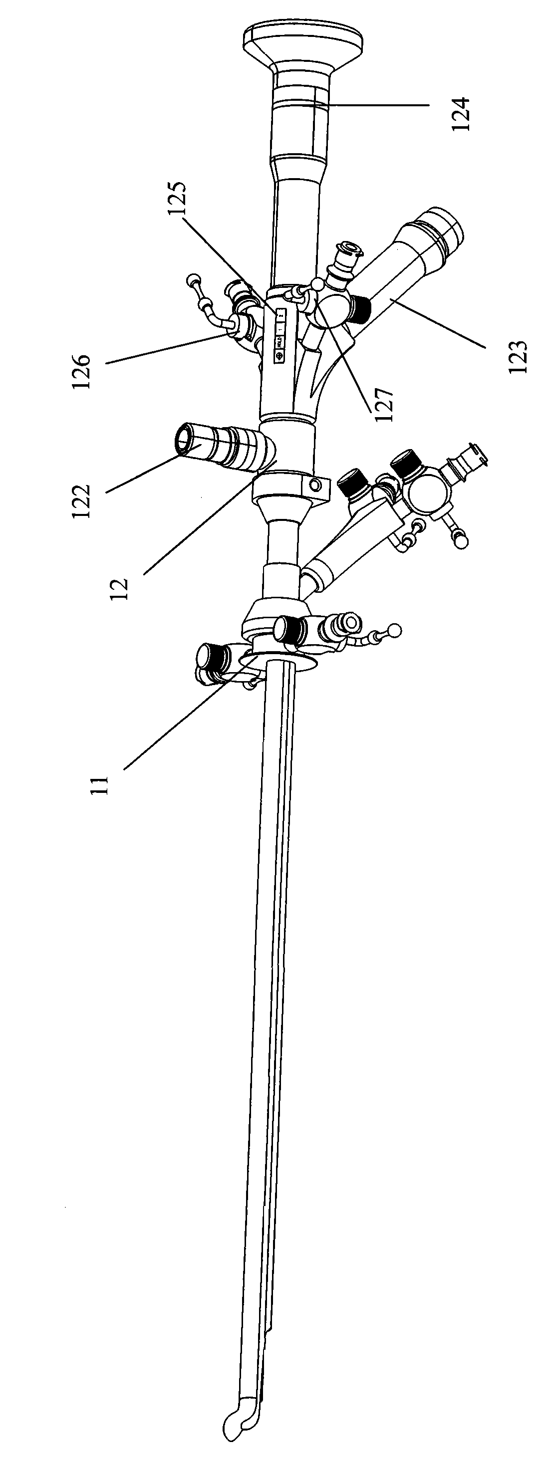
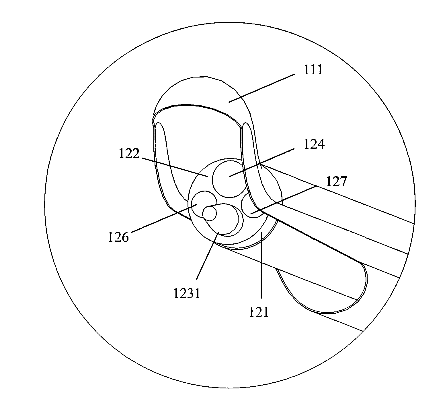
[0021] Such as figure 2 , combined with image 3 Shown is a schematic view of the integrated rigid ultrasonic cystoscope 1 in the present invention. The integrated rigid ultrasonic cystoscope 1 is divided into two parts: a sheath tube part 11 and a main endoscope part 12 . The top end of the sheath tube end 111 is designed to be blunt to prevent damage to the urethra. The length of the sheath tube end 111 is 180-220 mm, the shape of the sheath tube end 111 is circular or non-circular, and its maximum diameter is 10 mm. The main body endoscope part 12 of the integrated rigid ultrasonic...


PUM

PropertyMeasurementUnit
Outer diameteraaaaaaaaaa
Lengthaaaaaaaaaa
Diameteraaaaaaaaaa

Abstract

The invention belongs to the field of medical devices and particularly discloses an integrated rigid ultrasonic cystoscope system which comprises a rigid cystoscope and a cold light source host and a camera host connected with the rigid cystoscope, wherein the rigid cystoscope comprises a main body endoscope part and a cystoscope sheath tube part connected with the main body endoscope part, a micro-ultrasonic probe part, a light guide optical fiber and a device channel outlet are arranged at a rigid endoscope end part, and the micro-ultrasonic probe part is directly integrated at the rigid endoscope end part, thereby designing the rigid cystoscope into the integrated rigid ultrasonic cystoscope. The integrated rigid ultrasonic cystoscope system is placed into the bladder through the urethra of a patient, and a doctor operates a control unit of a cystoscope body for starting the micro-ultrasonic probe part of an endoscope, thereby carrying out annular scanning or linear scanning on the bladder cavity, displaying the scanning result in a monitor, carrying out three-dimensional reconstruction on the bladder cavity, knowing the situation of the bladder cavity and helping the doctor made judgments. The integrated design of the integrated rigid ultrasonic cystoscope leads the micro-ultrasonic probe part to be difficult to be damaged and facilitate the operation of the doctor; furthermore, an endoscope image and an ultrasonic image are easier to be obtained synchronously, thereby improving the image quality of the lesion area and greatly improving the accuracy of diagnosis.

Description

technical field [0001] The invention belongs to the field of medical equipment, and in particular relates to a core medical tool for performing urological surgery in modern medicine. Background technique [0002] At present, cystoscopy and cystoscopy technology have been widely used clinically, and are important tools for diagnosis and treatment of diseases in urology. However, the existing cystoscopy used for the diagnosis and treatment of bladder diseases can determine the lesion of the bladder, but cannot understand the layers of the bladder and its surroundings. Changes in organs have the potential for insufficient diagnostic accuracy. Micro-ultrasound technology is introduced into the field of urology surgery. Micro-ultrasound probes can be used to perform circular or linear scans to check the bladder wall and surrounding organs. [0003] Micro-ultrasound technology has been widely used in various fields of gastroenterology. Ultrasonic scanning of the ward through the m...

Claims


Application Information

Patent Timeline
IPC IPC(8): A61B1/317A61B1/018A61B1/07A61B8/00
Inventor 乔铁
Owner 广州市番禺区胆囊病研究所
Why Eureka
Try Eureka