

Method for distinguishing fungi and bacteria
A technology for bacteria and fungi, applied in biochemical equipment and methods, microbial determination/inspection, fluorescence/phosphorescence, etc., can solve the problems of complicated preparation, high cost, etc., to reduce drug resistance, simple sample processing, and fast processing. Effect
Active Publication Date: 2010-09-15
INST OF CHEM CHINESE ACAD OF SCI
View PDF4 Cites 4 Cited by
- Summary
- Abstract
- Description
- Claims
- Application Information
AI Technical Summary
Problems solved by technology
Although the existing detection methods have certain sensitivity and selectivity, the shortcomings of high cost, complicated preparation, long-term processing and special requirements for instruments limit the large-scale application of these methods
Method used
View moreImage
Smart ImageViewing Examples

Examples
Embodiment 1
Embodiment 2
Embodiment 3
Login to View More PUM
 Abstract
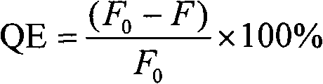
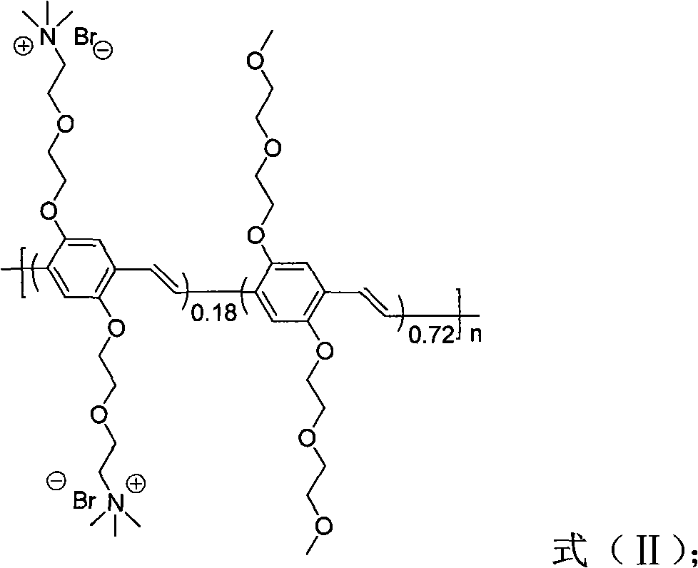
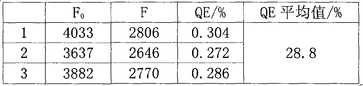
The invention discloses a method for distinguishing fungi and bacteria, which comprises the following steps of: detecting whether the bacteria to be detected can induce the compounds expressed by the formulas (I and II) to cause fluorescence resonance energy transfer; if the bacteria to be detected can induce the compounds expressed by the formulas (I and II) to cause fluorescence resonance energy transfer, determining that the bacteria to be detected is bacteria; and if the bacteria to be detected cannot induce the compounds expressed by the formulas (I and II) to cause fluorescence resonance energy transfer, determining that the bacteria to be detected is fungi. The method has the advantages of high speed, visibility and convenience and the like. The method can be used for distinguishing the types of the infectious pathogenic bacteria, instructing the administration of the doctors aiming at different patients, improving the therapeutic efficiency and reducing the occurrence of drug resistance caused by incorrect use of antibiotic. The method also has application value in aspects of food safety, environmental monitoring and the like.
Description
technical field The present invention relates to a method for differentiating fungi and bacteria. Background technique The detection of pathogenic bacteria is of great significance in the fields of medical treatment, food safety and environmental science. Since deep fungal infections are occult and have no characteristic clinical manifestations, the quick distinction between fungi and bacteria has important guiding significance for physicians to administer more targeted drugs clinically. In deep fungal infections, Candida albicans is the most common pathogen, accounting for about 40% of all types of fungi; while in blood-borne infections caused by bacteria, Escherichia coli is the most common type of bacteria, among which E.coli O157:H7 occupies a considerable proportion in various bacterial infections. At present, the traditional methods for the detection of pathogenic bacteria mainly include microbial culture methods, membrane filtration methods, and biochemical detecti...
Claims
Login to View More Application Information
Patent Timeline
 IPC IPC(8): C08G61/02C12Q1/04G01N21/64
Inventor王树杨琼朱春雷
OwnerINST OF CHEM CHINESE ACAD OF SCI