Multi-mode imaging system for observing cerebral cortex functions of moving animals

A multimodal imaging, animal brain technology for biomedical imaging applications

Active Publication Date: 2010-12-22
HUAZHONG UNIV OF SCI & TECH
View PDF6 Cites 17 Cited by
  • Summary
  • Abstract
  • Description
  • Claims
  • Application Information

AI Technical Summary

Problems solved by technology

[0005] The purpose of the present invention is to provide a multi-mode imaging system that can be used for the observation of cerebral cortex function of awake animals. Contrast Imaging and Fluorescence Imaging Issues

Method used


Image

  • Multi-mode imaging system for observing cerebral cortex functions of moving animals
  • Multi-mode imaging system for observing cerebral cortex functions of moving animals
  • Multi-mode imaging system for observing cerebral cortex functions of moving animals

Examples


Embodiment Construction

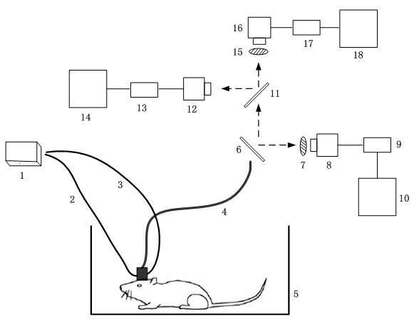
[0024] figure 1 It is a structural diagram of a multi-mode imaging system for observing the cerebral cortex of an active animal according to the present invention, specifically including:

[0025] Light source device 1, light transmission fiber 2, light transmission fiber 3, image transmission fiber 4, animal activity room 5, beam splitter 6, filter 7, charge coupler 8, image acquisition card 9, computer 10, beam splitter 11, A charge coupler 12, an image acquisition card 13, a computer 14, an optical filter 15, a charge coupler 16, an image acquisition card 17, and a computer 18.

[0026] One end of the light transmitting optical fiber 2 and the light transmitting optical fiber 3 is connected to the light source device 1, and the other end is fixed on the head of the animal. One end of the image transmission fiber 4 is fixed on the head of the animal, and the other end is fixed in front of the beam splitter 6 . A filter 7 and a charge coupler 8 are placed on the right side ...


PUM


Abstract

The invention discloses a multi-mode imaging system for observing cerebral cortex functions of moving animals, comprising a light source device, an image transferring optical fiber, a light transferring optical fiber, a spectroscope, an intrinsic signal imaging part, a laser speckle contrast imaging part, a fluorescence Imaging part, an animal activity room, an image acquisition card, a computer, wherein the light source device generates three light sources needed by an imaging mode, the image transferring optical fiber is used for transferring the cerebral cortex images to a spectroscope when the animals moves around freely; the light transferring optical fiber transfers the light of the light source to the cerebral cortex of the aminals; the spectroscope divides imaging light beam transferred by the image transferring optical fiber into three beams and carries out intrinsic signal imaging, laser speckle contrast imaging and fluorescence Imaging; the intrinsic signal imaging part is used for the intrinsic signal imaging; the laser speckle contrast imaging part for laser speckle contrast imaging; the fluorescence Imaging part is used for fluorescence Imaging; the animal activity room is used for the experiment animals to move freely; the image acquisition card is used for image acquisition and the computer for system control and image receiving and processing.In the invention, optical fiber light and image transferring can be used to multi-mode imaging when the animals are conscious and active and the invention comprises intrinsic signal imaging, laser speckle contrast imaging and fluorescence Imaging.

Description

technical field [0001] The present invention relates to biomedical imaging technology, especially the combination of endogenous signal optical imaging technology, laser speckle contrast imaging technology and fluorescence imaging technology. pattern imaging. Background technique [0002] Brain function optical imaging has always been an extremely important research method in the field of neuroscience research. At present, the optical imaging methods of brain function widely carried out at home and abroad include various imaging methods such as fluorescence imaging, optical imaging of endogenous signals, near-infrared light imaging, and laser speckle imaging. Most of these imaging methods are applied to small animals that have been anesthetized and fixed on the bench. Although optical imaging systems based on anesthetized small animals have been widely used, and can provide a very stable platform for scientific research on brain function, the physiological responses of ani...

Claims


Application Information

Patent Timeline
Patent Type & Authority Applications(China)
IPC IPC(8): A61B5/00
Inventor 李鹏程骆清铭尹翠刘睿孙小丽
Owner HUAZHONG UNIV OF SCI & TECH
Why Eureka
Try Eureka