Novel three-dimensional electronic choledochoscope system and use method thereof

A three-dimensional and choledochoscope technology, applied in esophagoscope, urethroscope, gastroscope, etc., can solve the problems of not being able to reflect the three-dimensional panorama of the operation area three-dimensionally, the limitation of the cure rate of the disease during the operation, and the single angle of the image.

Active Publication Date: 2012-05-30
GUANGZHOU BAODAN MEDICAL INSTR TECH
View PDF0 Cites 0 Cited by
  • Summary
  • Abstract
  • Description
  • Claims
  • Application Information

AI Technical Summary

Problems solved by technology

[0004] Some stereoscopic endoscopes use a binocular structure, and the front end of the endoscope can be one lens or two lenses. The image of the object is output through the two eyepieces. The doctor can observe through the binoculars similar to the human eye. The three-dimensional image of the object can also be connected to a special processing host and the display, and the three-dimensional image can be displayed on the display through the processing of the processing host, but this image is also a single angle, which has certain limitations
Although the binocular stereoscopic endoscope can obtain the stereoscopic feeling similar to the human eye observing objects, but due to the limitation of the human body cavity, it cannot stereoscopically reflect the stereoscopic picture of the entire operation area, so doctors are using the current endoscope for surgery. At the same time, it must be restricted by vision, which has certain restrictions on the development of surgery and the improvement of the cure rate of the disease.
[0005] In the prior art, the concept of CCD array has not been combined with flexible choledochoscope. Therefore, in order to obtain a clear three-dimensional image in the bile duct, an endoscopic technology combining multi-CCD array technology and fiber choledochoscope is designed. imminent

Method used


Image

  • Novel three-dimensional electronic choledochoscope system and use method thereof
  • Novel three-dimensional electronic choledochoscope system and use method thereof
  • Novel three-dimensional electronic choledochoscope system and use method thereof

Examples


Embodiment Construction

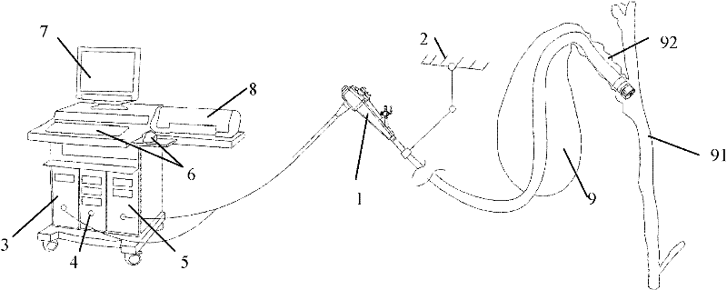
[0029] like figure 1 As shown, the novel three-dimensional electronic choledochoscope system of the present invention comprises a soft electronic choledochoscope 1, an external fixed bracket 2, and a processing host 3 connected thereto, a light source host 5, and a workstation assembly. The workstation assembly includes a workstation host 4, a control Components 6 (keyboard and mouse, etc.), monitor 7 and external devices 8 (external storage, printer, etc.), etc. The workstation component is connected to the processing host through a data line, and the function of the workstation component is to display the three-dimensional image output by the processing host 3, analyze and store data, and print related materials. The core part of the processing host 3 adopts a high-speed central processing unit and a high-performance graphics card for receiving and processing the data packets composed of the image information returned by the soft electronic choledochoscope 1 and the data of ...


PUM


Abstract

The invention belongs to medical instruments, and in particular relates to a novel three-dimensional electronic choledochoscope system for reconfiguring a three-dimensional image by using a multi-CCD (Charge Coupled Device) array, which comprises a soft electronic choledochoscope, a processing host computer, a light source host computer and a work station assembly, wherein the processing host computer, the light source host computer and the work station assembly are connected with the soft electronic choledochoscope; the three-dimensional soft electronic cystoscope enters a gall bladder cavity through a small cut at the bottom of the gall bladder and can enter the biliary tract through a small cut on the bile duct wall, then turns into the bile duct through better flexibility and control,and then enters the common bile duct for detecting the condition of the common bile duct; the multi-CCD array module at the tip part of a choledochoscope working end part is used for finely scanning the common bile duct, even the intrahepatic bile duct to obtain a three-dimensional image of the choledochoscope cavity; and a doctor treats the gall bladder pathological change according to the three-dimensional image.

Description

technical field [0001] The invention belongs to medical equipment, and in particular relates to a novel three-dimensional electronic choledochoscope system which utilizes multiple CCD arrays to reconstruct stereo images. Background technique [0002] A CCD (Charge-Coupled Device, Charge-Coupled Device) is an important component that can be used in a stereo camera. It is a photosensitive semiconductor device on which the photosensitive unit converts the received light into electric charge, and the electric charge is proportional to the intensity of incident light. The technology of CCD image sensor is extremely mature, and it can be spliced ​​into arrays of any shape as required. In 1999, Fuji launched the super CCD technology. Under the same area and number of photosensitive units as the ordinary CCD, its resolution increased by 60%, the dynamic range increased by 130%, the color reproduction ability increased by 40%, and the energy consumption decreased by 40%. Improve th...

Claims


Application Information

Patent Timeline
Patent Type & Authority Patents(China)
IPC IPC(8): A61B1/313A61B1/273A61B1/05
CPCA61B1/307A61B1/00096A61B1/05
Inventor 乔铁
Owner GUANGZHOU BAODAN MEDICAL INSTR TECH
Why Eureka
Try Eureka