X-ray compound tomoscanning imaging system and method

A tomography and imaging system technology, which is applied to the computerized duplex tomography imaging system, can obtain high-resolution tomographic images in the imaging field, and can solve the problems of poor imaging quality, low imaging resolution, and poorly targeted CT images. , to achieve the effect of improving efficiency, improving resolution and reducing labor consumption

Inactive Publication Date: 2011-06-29
HOHAI UNIV
View PDF4 Cites 39 Cited by
  • Summary
  • Abstract
  • Description
  • Claims
  • Application Information

AI Technical Summary

Problems solved by technology

[0009] The purpose of the present invention is to overcome the shortcomings of low imaging resolution and poor imaging quality of existing CT imaging equipment, and at the same time, when using the technology of changing time for space, in order to solve the problem that the common super-resolution method is not very specific to the characteristics of CT images question

Method used


Image

  • X-ray compound tomoscanning imaging system and method
  • X-ray compound tomoscanning imaging system and method
  • X-ray compound tomoscanning imaging system and method

Examples


Embodiment Construction

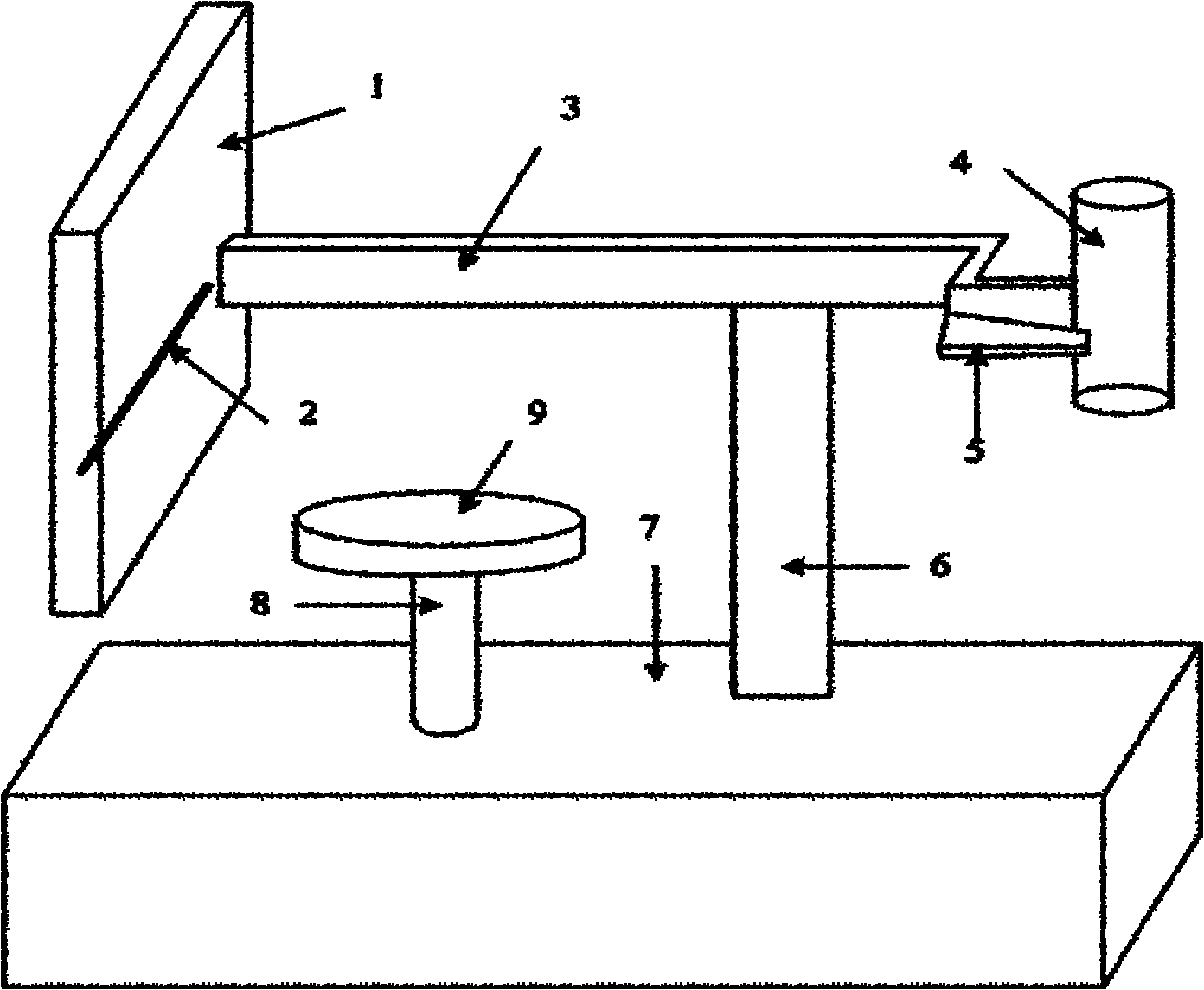
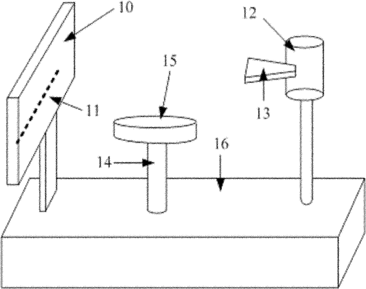
[0031] Such as image 3 As shown, the present invention uses two connecting columns to respectively fix and support the film box 10 and the X-ray tube 12 at the two ends of the base 16, and the X-ray tube 12 is equipped with a beam device for guiding the direction and shape of the rays. 13. The film box 10 has a built-in X-ray linear array detector 11 that receives X-rays, and the X-rays guided by the light beam device 13 are aimed at the X-ray line array detector 11. The film box 10 and X-ray In the middle of the ray tube 12 is a table 15 that can be lifted and rotated for placing and fixing the measured object. The table 15 is connected to the base 16 through the lifting shaft 14. and driven by the rotary control device. When objects need to be scanned, the X-ray tube 12, the X-ray linear array detector 11 and the objects on the table top 15 should be adjusted to the horizontal line of the same height by the lifting shaft. When an object is to be scanned with X-rays, the t...


PUM


Abstract

The invention discloses an X-ray compound tomoscanning imaging system and method. The whole imaging system consists of a mechanical device (comprising a shooting box, an X-ray linear array detector, an X-ray bulb tube, a light concentrator, a lifting rotating shaft and a table surface) and a monitoring feedback control system. The method comprises the steps of firstly setting the frame number N of a sequence image needed by super-resolution reconstruction; performing total body X-ray scan on a monitored object to obtain an X-ray planar image; horizontally moving the small distance of a sub-detection unit by the detector until an image sequence of displacements of N frames of sub-pixels is obtained; acquiring an X-ray planar image with a high resolution in a super-resolution reconstructionimage sequence, and finding out an interested point in the image; rotationally tomoscanning the interested point in the fault which the interested point is positioned at so as to reconstruct a computed tomography (CT) image; horizontally moving the small distance of the sub-detection unit by the detector until the image sequence of displacements of N frames of sub-pixels displacement is obtained;and super-resolution reconstructing the image sequence to obtaining the CT image with a high resolution in the super-resolution reconstruction image sequence.

Description

technical field [0001] The invention relates to a computer multiple tomography imaging system, in particular to an imaging method capable of obtaining high-resolution tomographic images, belonging to the field of CT signal and image processing. Background technique [0002] X-ray is a kind of electromagnetic radiation with very short wavelength. It has high penetrating power and can penetrate many substances that are opaque to visible light. Therefore, it is widely used in the fields of medical diagnosis and industrial detection. The common X-ray imaging is accomplished by aligning the X-ray tube with the light beam device and the linear detector composed of the detection and sensing unit to scan the object under test in synchronous motion. X-rays have different penetration capabilities to different densities, structures and components inside the measured object, so the energy induced by the detector through the measured object is also different. When the ray transceiver co...

Claims


Application Information

Patent Timeline
Patent Type & Authority Applications(China)
IPC IPC(8): A61B6/03G01N23/04
Inventor 徐枫徐立中王慧斌王鑫张学武石爱业张振张家华丁晓峰郭锐
Owner HOHAI UNIV
Why Eureka
Try Eureka