

Direct monitoring method of mast cell degranulation
A mast cell, degranulation technique, applied in the field of determination of substances that cause anaphylactoid reactions
- Summary
- Abstract
- Description
- Claims
- Application Information
AI Technical Summary
Problems solved by technology
Method used
Image

Examples
Embodiment 1
[0087] Embodiment 1, measuring method
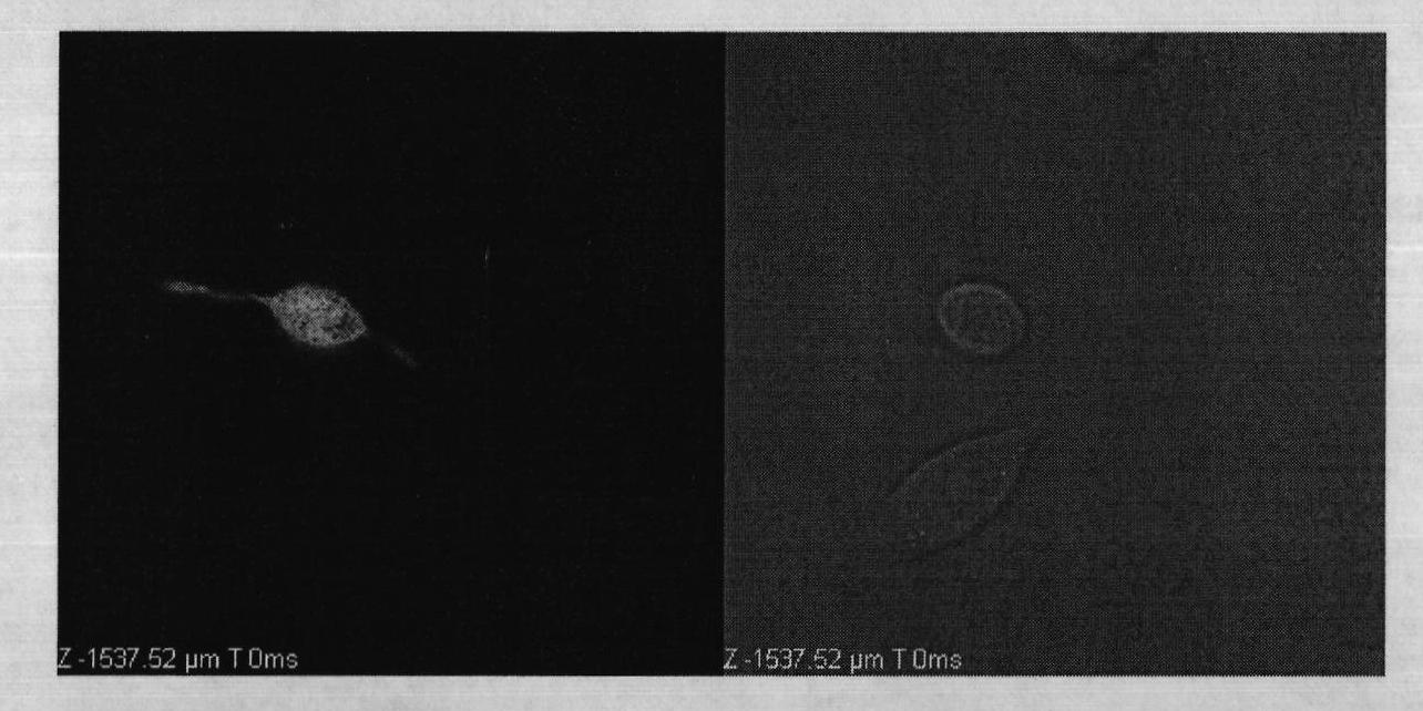
[0088]Step 1, the RBL-2H3 cells transfected with CD63-GFP plasmid were cultured with MEM medium (MEM medium: MEM powder 9.4g, L-glutamine 0.292g, NaHCO 3 Add 1000ml of three-distilled water to 2.2g, add 10% fetal bovine serum, 100,000 units of penicillin and streptomycin when used), after three passages, inoculate the cells into a confocal culture dish (diameter 3cm, glass bottom thickness 0.17μm) at 37°C After culturing in the incubator for 24 hours;
[0089] Step 2, replace the culture medium with Tyrode's solution (preparation method: NaCl 400mg, KCl 100mg, MgCl 2 50mg, NaHCO 3 500mg, NaH 2 PO 4 32.5 mg, CaCl 2 100g, add distilled water to 1000ml, add glucose 1.0g before use), incubate at 37°C for 30min;
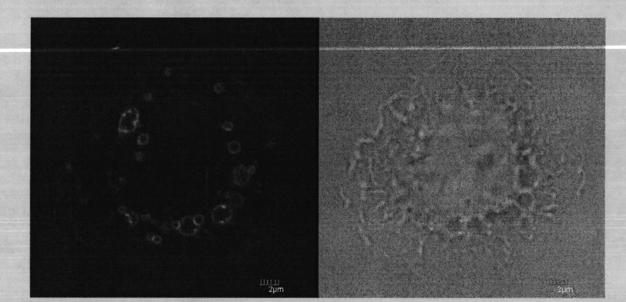
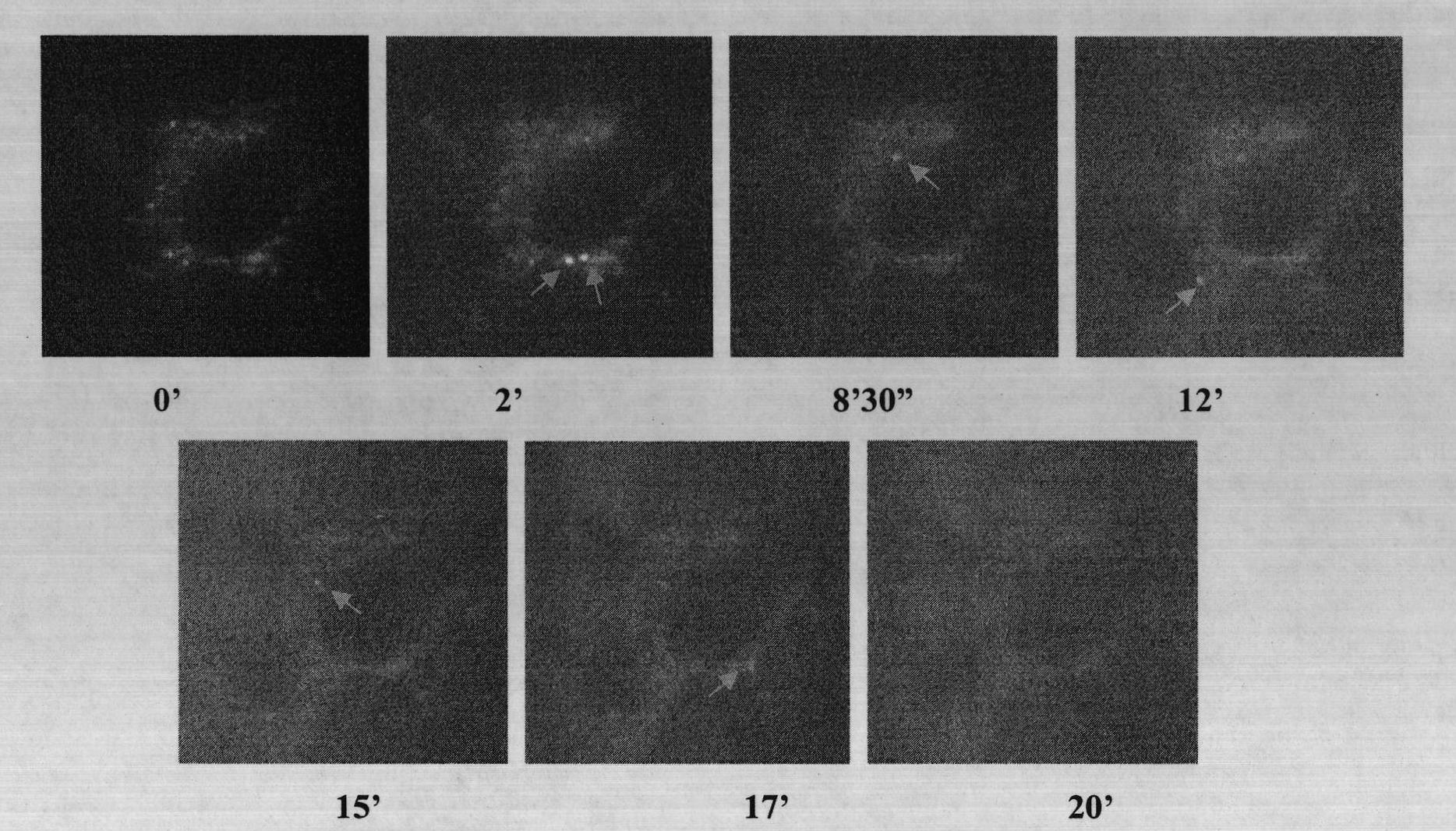
[0090] Step 3, after adding the test drug, move to a laser scanning confocal microscope for observation under a 100X oil lens. After selecting a field of view with good cell growth and no overlap with each other, images w...
Embodiment 2
[0097] Embodiment 2, allergic substance assay method
[0098] Step 1, the RBL-2H3 cells transfected with CD63-GFP plasmid were cultured with MEM medium (MEM medium: MEM powder 9.4g, L-glutamine 0.292g, NaHCO 3 Add 1000ml of three-distilled water to 2.2g, add 10% fetal bovine serum, 100,000 units of penicillin and streptomycin when used), after three passages, inoculate the cells into a confocal culture dish (diameter 3cm, glass bottom thickness 0.17μm) at 37°C After culturing in the incubator for 24 hours;
[0099] Step 2, replace the culture medium with Tyrode's solution (preparation method: NaCl 400mg, KCl 100mg, MgCl 2 50mg, NaHCO 3 500mg, NaH 2 PO 4 32.5 mg, CaCl 2 100g, add distilled water to 1000ml, add glucose 1.0g before use), incubate at 37°C for 30min;
[0100] Step 3, after adding the suspected allergic substance to be tested, move to a laser scanning confocal microscope for observation under a 100X oil lens. After selecting a field of view with good cel...
Embodiment 3
[0107] Embodiment 3, Chinese medicine injection allergen determination method
[0108] Step 1, the RBL-2H3 cells transfected with CD63-GFP plasmid were cultured with MEM medium (MEM medium: MEM powder 9.4g, L-glutamine 0.292g, NaHCO 3 Add 1000ml of three-distilled water to 2.2g, add 10% fetal bovine serum, 100,000 units of penicillin and streptomycin when used), after three passages, inoculate the cells into a confocal culture dish (diameter 3cm, glass bottom thickness 0.17μm) at 37°C After culturing in the incubator for 24 hours;
[0109] Step 2, replace the culture medium with Tyrode's solution (preparation method: NaCl 400mg, KCl 100mg, MgCl 2 50mg, NaHCO 3 500mg, NaH 2 PO 4 32.5 mg, CaCl 2 100g, add distilled water to 1000ml, add glucose 1.0g before use), incubate at 37°C for 30min;
[0110] Step 3, after adding the traditional Chinese medicine injection, move to a laser scanning confocal microscope for observation under a 100X oil lens. After selecting a field...
PUM
 Abstract
Description
Claims
Application Information