A method to synthesize α-galactoside epitopes on the surface of tumor cells and activate immune cells

A technology of tumor cells and galactoside, which is applied in the direction of tumor/cancer cells, animal cells, vertebrate cells, etc., and can solve problems such as the inability of immunotherapy technology

Inactive Publication Date: 2011-12-21
INNER MONGOLIA MEDICAL COLLEGE
View PDF1 Cites 1 Cited by
  • Summary
  • Abstract
  • Description
  • Claims
  • Application Information

AI Technical Summary

Problems solved by technology

In order to synthesize α-galactoside epitopes on the surface of human tumor cell membranes, the immunotherapy techniques described in the above literatures all need to use live tumor samples, and for those patients who did not keep live tumor samples at that time, this immunotherapy technique is unable to proceed

Method used


Image

  • A method to synthesize α-galactoside epitopes on the surface of tumor cells and activate immune cells
  • A method to synthesize α-galactoside epitopes on the surface of tumor cells and activate immune cells
  • A method to synthesize α-galactoside epitopes on the surface of tumor cells and activate immune cells

Examples


Embodiment 1

[0015] Example 1. Isolation of Tumor Cells from Formalin-Fixed Human Tumor Tissue

[0016] The tumor specimens were cut into tissue pieces smaller than 1cm, soaked in a large amount of cold saline, and shaken on the shaker in the cold storage for at least 6 hours, during which the saline was changed at least three times. Carefully remove all connective tissue around the tumor tissue under a microscope, free tumor cells with a thin curved needle, filter with a cell sieve, remove large cell clumps and connective tissue, and wash the free tumor cells three times with cold saline.

Embodiment 2

[0017] Example 2. Isolation of tumor cells from paraffin-embedded tumor tissue

[0018] Use a scalpel to remove all the paraffin around the tumor specimen as much as possible, prepare a series of Histo-clear solutions, 3 containers with covers, each 100ml, and place them on a shaker in a fume hood; place the treated tumor specimens on the above-mentioned In one container, after all the paraffin is dissolved, transfer to the second and third containers to further remove the paraffin; transfer the above-mentioned dewaxed specimens to the following four alcohol series containers (100%, 50%, 20 % and normal saline), each on a shaker for more than one hour; free tumor cells with a thin curved needle, filter with a cell sieve, remove large cell clumps and connective tissue, and wash free tumor cells three times with cold normal saline.

Embodiment 3

[0019] Example 3. Synthesis of α-Gal epitopes on the surface of fixed tumor cells

[0020] 3.1 Configure enzyme buffer 1ml (0.1M MES (PH 6.2), 25mM MnCl 2 and 1mM UDP-gal). Resuspend the above isolated tumor cells with 1 ml enzyme buffer. Then add 1mU neuraminidase and 5U recombinant bovine α-1,3-GT to the system, incubate at 37°C for 30 minutes, take out 200μl for flow cytometry:

[0021] Negative control group: no sialidase (neuraminidase), no GT

[0022] Experimental group: with sialidase, with GT

[0023] After the incubation, wash twice with normal saline containing 1 mM EDTA and saline without EDTA respectively (1500 rpm, centrifuged for 10 minutes).

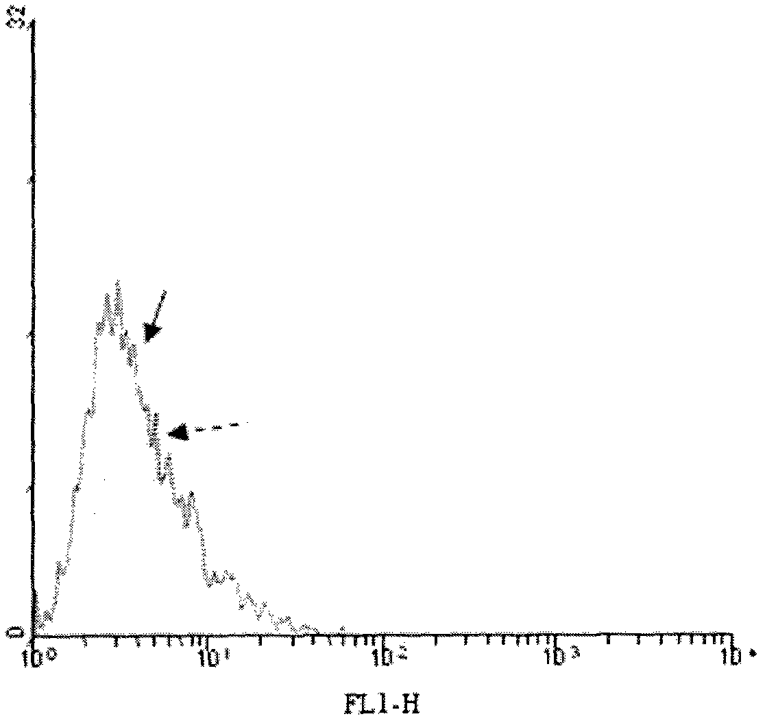
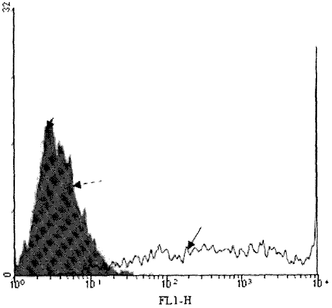
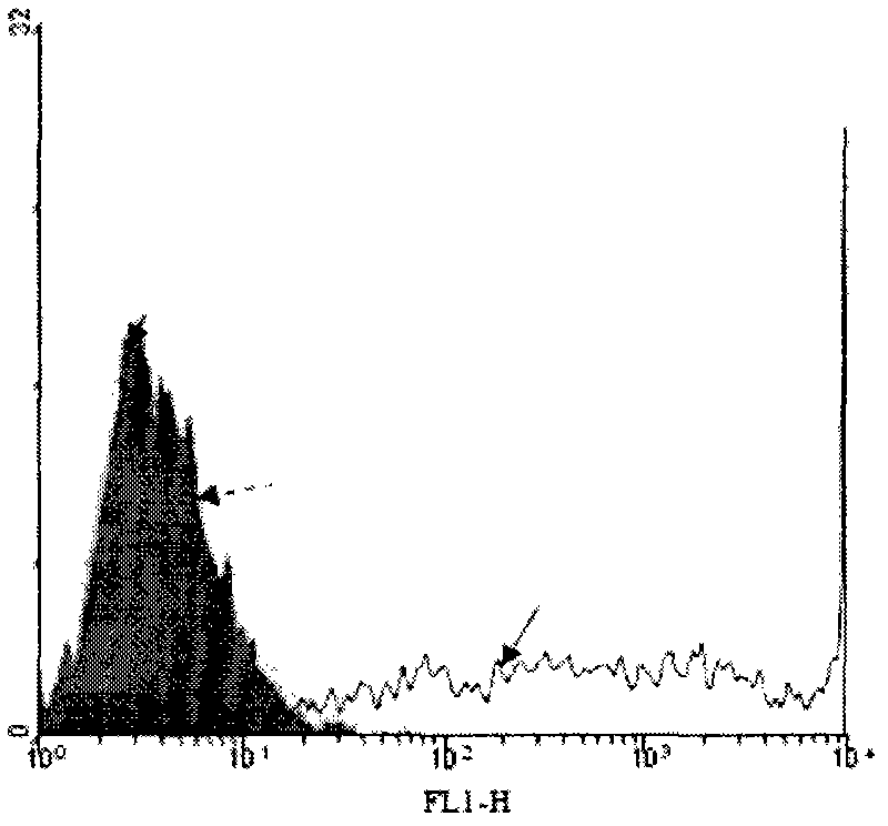
[0024] 3.2 Determination of α-Gal epitope synthesis on the surface of tumor cell membrane by flow cytometry

[0025] Firstly, 1 ml of human serum (containing natural anti-α-Gal epitope antibody) was diluted 1:5 with 1% BSA saline, mixed with cells, and incubated on ice for 60 minutes. Then wash twice with 1% BSA phys...


PUM


Abstract

The present invention relates to a method for synthesizing α-galactoside epitope (α-Gal) on the surface of tumor cells and activating immune cells, using old, formalin-fixed or paraffin-embedded tumor specimens, through α-1,3-galactoside transferase catalyzes the synthesis of galactoside (α-Gal) epitopes on tumor cell membranes, enhances tumor antigenicity, and uses it as a vaccine. The α-Gal epitope on these tumor cell membranes can be phagocytized by antigen-presenting cells (APC) in vitro, activate T/CIK cells, and rapidly expand tumor-specific T/CIK cells in vitro. tumor cells.

Description

technical field [0001] The present invention relates to a method for synthesizing α-galactoside epitope (α-Gal) on the surface of tumor cells and activating immune cells. Specifically, the present invention utilizes old, formalin-fixed or paraffin-coated Buried tumor samples, catalyzed by α-1,3-galactosidyl transferase (GT), synthesize galactoside (α-Gal) epitopes on tumor cell membranes, enhance tumor antigenicity, and use them as vaccines. The α-Gal epitope on these tumor cell membranes can be phagocytized by APC cells in vitro, activate T cells and CIK cells, and at the same time rapidly expand tumor-specific T cells and CIK cells in vitro, and these expanded immune cells can specifically kill tumor cells in vivo. Background technique [0002] The basic principle of specific immunotherapy for tumors is to induce effective immune responses against tumor associated antigens (TAA), thereby killing residual tumor cells. However, the use of autologous tumor cells as a tumor ...

Claims


Application Information

Patent Timeline
Patent Type & Authority Applications(China)
IPC IPC(8): C12N5/09C12N5/0783
Inventor 云升邱英
Owner INNER MONGOLIA MEDICAL COLLEGE
Why Eureka
Try Eureka