Method for measuring distribution of human cell drug in animal body

A technology of human cells and animal bodies, applied in the biological field, can solve the problems of inability to detect the content of monkey human cells, inability to distinguish human and monkey chromosomal DNA, and narrow application range.

Inactive Publication Date: 2012-05-16
北京昭衍新药研究中心股份有限公司
View PDF0 Cites 2 Cited by
  • Summary
  • Abstract
  • Description
  • Claims
  • Application Information

AI Technical Summary

Problems solved by technology

This method can be used to study the distribution of human cell drugs in animals with low homology to humans, such as mice and rats. However, due to the high homology of human and monkey gene sequences, the housekeeping gene sequences are basically the same. It is impossible to distinguish between the chromosomal DNA of humans and monkeys, and it is also impossible to detect the content of human cells in monkeys
Therefore, this method is currently only used in the study of the distribution of human cells in rodents, and its application range is relatively narrow.

Method used


Image

  • Method for measuring distribution of human cell drug in animal body
  • Method for measuring distribution of human cell drug in animal body
  • Method for measuring distribution of human cell drug in animal body

Examples


Embodiment 1

[0058] (1) According to the human specific gene UCMA, select the PCR amplification region (2281-3618), namely SEQ ID NO. 1, and design PCR amplification primers. The primer sequence is as follows.

[0059] Upstream primer: catttagcctgaagtgcctt

[0060] Downstream primer: ccttgtttctcccagcacctctg

[0061] (2) Using conventional tissue total DNA extraction kit (TAKARA, DV811A), extract genomic DNA from whole blood of healthy people, amplify the target gene fragment according to the following reaction conditions, PCR amplification reaction kit (Tiangen, ET101-01 ).

[0062] Reaction system: 0.1ug template, 500nM upstream and downstream primers, add water to 50ul,

[0063] Reaction conditions: 95°C pre-denaturation 10min; 95°C denaturation 15sec, 50°C annealing 15sec, 72°C extension 90sec, 30 cycles; 72°C extension 5min;

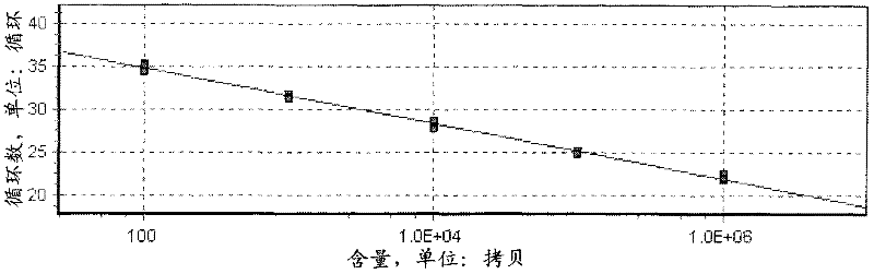
[0064] (3) Operate according to the pMD19-T (TAKARA, VT202-01) instruction, connect the target gene fragment into the pMD19-T vector to construct a standard plasmid, and d...

Embodiment 2

[0078] (1) According to the human specific gene UCMA, select the PCR amplification region (10081-10740), namely SEQ ID NO. 3, and design PCR amplification primers. The primer sequence is as follows.

[0079] Upstream primer: ggctcgagcaatcctcccaa

[0080] Downstream primer: ctgcaccccagcctgggtga

[0081] (2) Using conventional tissue total DNA extraction kit (TAKARA, DV811A), extract genomic DNA from whole blood of healthy people, amplify the target gene fragment according to the following reaction conditions, PCR amplification reaction kit (Tiangen, ET101-01 ).

[0082] Reaction system: 0.1ug template, 200nM upstream and downstream primers, add water to 50ul,

[0083] Reaction conditions: 95°C pre-denaturation 10min; 95°C denaturation 15sec, 52°C annealing 15sec, 72°C extension 90sec, 35 cycles; 72°C extension 7min.

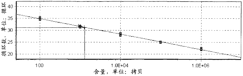
[0084] (3) Operate according to the pMD19-T (TAKARA, VT202-01) instruction, connect the target gene fragment into the pMD19-T vector to construct a standard plasmid, and de...

Embodiment 3

[0098] (1) According to the human specific gene UCMA, select the PCR amplification region (11281-11880), namely SEQ ID NO. 5, and design PCR amplification primers. The primer sequence is as follows.

[0099] Upstream primer: gtttcaccatgttggccaagtta

[0100] Downstream primer: tgaccttcgcccatctgggc

[0101] (2) Using conventional tissue total DNA extraction kit, extract genomic DNA from whole blood of healthy people, amplify target gene fragments according to the following reaction conditions, PCR amplification reaction kit (Tiangen, ET101-01);

[0102] Reaction system: 0.1ug template, 300nM upstream and downstream primers, add water to 50ul,

[0103] Reaction conditions: pre-denaturation at 94°C for 10 minutes; denaturation at 94°C for 15 seconds, annealing at 55°C for 15 seconds, extension at 72°C for 90 seconds, 30 cycles; extension at 72°C for 7 minutes.

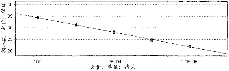
[0104] (3) Operate according to the pMD19-T (TAKARA, VT202-01) instruction, connect the target gene fragment into the pMD19-T vecto...


PUM


Abstract

The invention provides a method for measuring distribution of a human cell drug in an animal body, comprising the following steps of: (1) selecting a PCR(polymerase chain reaction) amplification region according to a human specific gene and designing a PCR amplification primer; (2) extracting human genome DNA(deoxyribonucleic acid), carrying out PCR amplification on a target gene fragment; (3) connecting the amplified fragment with a T vector to construct a standard plasmid; (4) establishing a standard curve by using the standard plasmid; (5) after the human cell drug is applied to an animal, collecting tissues, extracting a total DNA sample to carry out quantitative PCR reaction; and (6) calculating the contents of human genome DNA in the total DNA sample of each tissue by the standard curve, thereby calculating the contents of the human cell drug in the tissues. The measuring method provided by the invention has simple experimental processes and accurate and reliable experimental results.

Description

Technical field [0001] The invention relates to the field of biotechnology, in particular to a method for determining the distribution of human cell drugs in animals. Background technique [0002] With the development of human somatic cell research, the role of various human cells (especially mesenchymal stem cells) in the treatment of various human diseases has gradually attracted attention, such as liver damage, immune damage, nerve damage, blood For the treatment of diseases, the preclinical safety evaluation of this new type of therapeutic drug is particularly important. It is urgent to study the distribution of human cell drugs in animals after the drug. [0003] At present, there are two main methods to study the distribution of human cell drugs in animals: one method is to modify human cell drugs (Ju Shenghong, Teng Gaojun, Mao Xi, etc., cord blood mesenchymal stem cell magnetic probe labeling and MR imaging Research, Chinese Journal of Radiology, 2005, 39: 101-106; Bos C, ...

Claims


Application Information

Patent Timeline
Patent Type & Authority Applications(China)
IPC IPC(8): C12Q1/68
Inventor 李洪贞左从林孙云霞龚兆龙冯宇霞
Owner 北京昭衍新药研究中心股份有限公司
Why Eureka
Try Eureka