

Sequence specific DNA binding by p53
a dna sequence and tumor suppressor technology, applied in the field of identifying dna sequences specifically bound by the tumor suppressor p53, to achieve the effect of suppressing neoplastic growth
- Summary
- Abstract
- Description
- Claims
- Application Information
AI Technical Summary
Benefits of technology
Problems solved by technology
Method used
Image

Examples
example 1
[0093]This example demonstrates the screening methods used to identify a p53 sequence-specific binding site.
[0094]In an attempt to identify a possible sequence-specific binding site, numerous cloned DNA sequences were screened using an immunoprecipitation technique. The immunoprecipitation assay for DNA-binding was modified from R. D. G. McKay, J. Mol. Biol. 145. 471 (1981). Binding reactions included 5 μl of vaccinia-infected cell lysate or purified baculovirus p53 preparations, 95 μl binding buffer (20 mM Tris pH 7.2, 100 mM NaCl, 10% glycerol, 1% NP-40, 5 mM EDTA), 6–20×104 dpm 32P-labelled DNA, 4 μl (0.4 μg) pAb421 and 4 μl (0.4 μg) pAb1801 anti-p53 purified monoclonal antibodies (Oncogene Science), at 4° C. for 30 min. 1.5 mg protein A-Sepharose (Sigma) and 12.5 μg poly-dIdC-poly-dIdC (Pharmacia), in 25 μl binding buffer, were added and the reactions rotated end-over-end at 4° C. for 30 min. The pellet was washed twice with binding buffer and the proteins digested with SDS-prot...
example 2
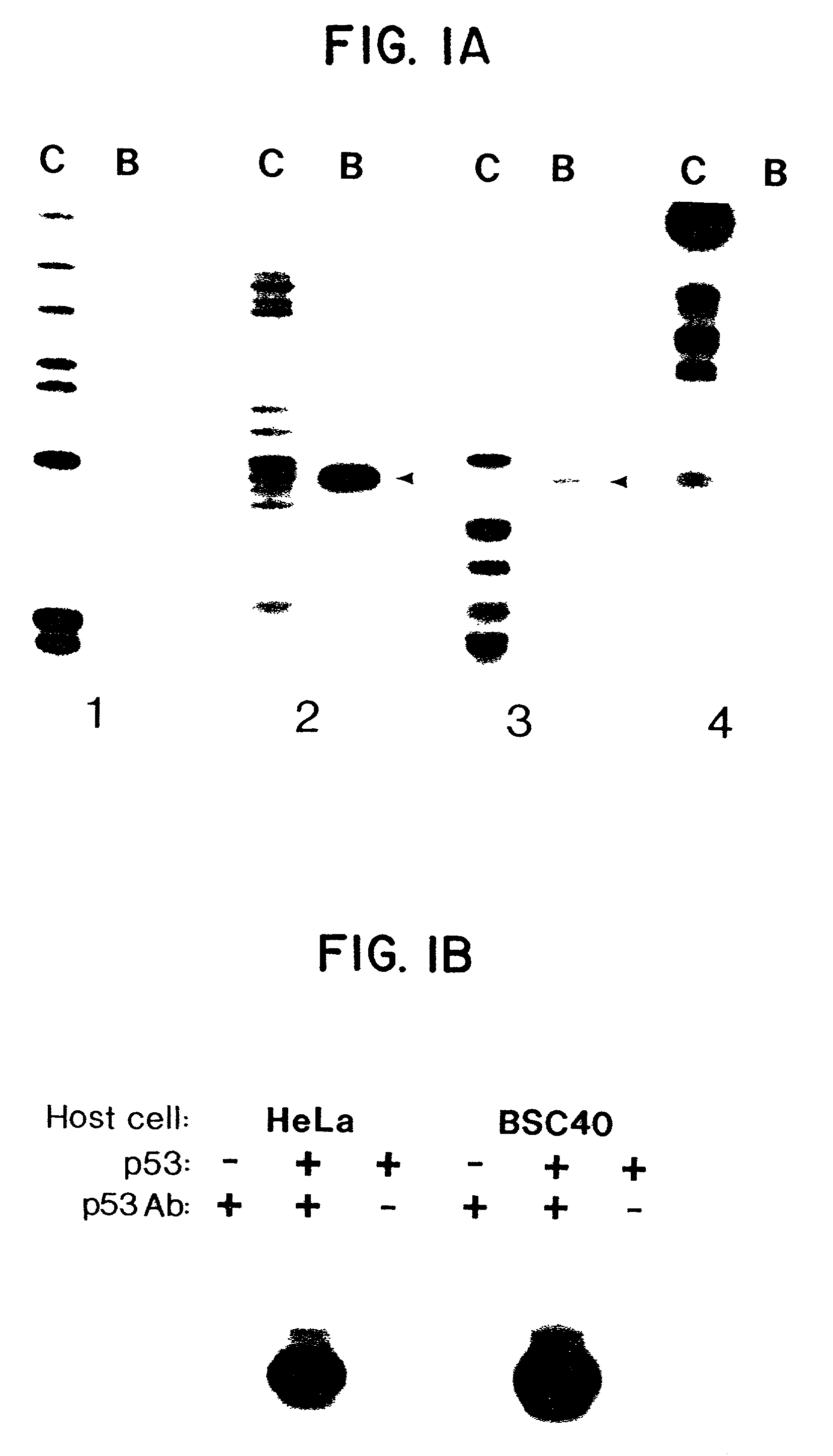
[0100]This example demonstrates that the immunoprecipitation of fragment A is dependent on both p53 protein and anti-p53 antibodies.
[0101]The immunoprecipitation assay was performed on fragment A as described in Example 1. Cell lysates were produced by infection with vaccinia virus that did or did not contain an insert of wild-type p53 DNA. Either anti-p53 antibodies were used or normal mouse IgG was used.
[0102]Lysates from cells infected with wild-type vaccinia virus (devoid of p53) were not able to specifically immunoprecipitate fragment A (FIG. 1B). Similarly, the detection of the precipitation of fragment A was dependent on the presence of anti-p53 antibodies (FIG. 1B). The binding was evident in lysates prepared from either human HeLa cells or monkey BSC40 cells infected with vaccinia virus and expressing wild-type p53 (FIG. 1B).
[0103]Affinity-purified baculovirus-produced wild-type p53 protein was substituted for the vaccinia-infected cell lysates in the immunoprecipitation as...
example 3
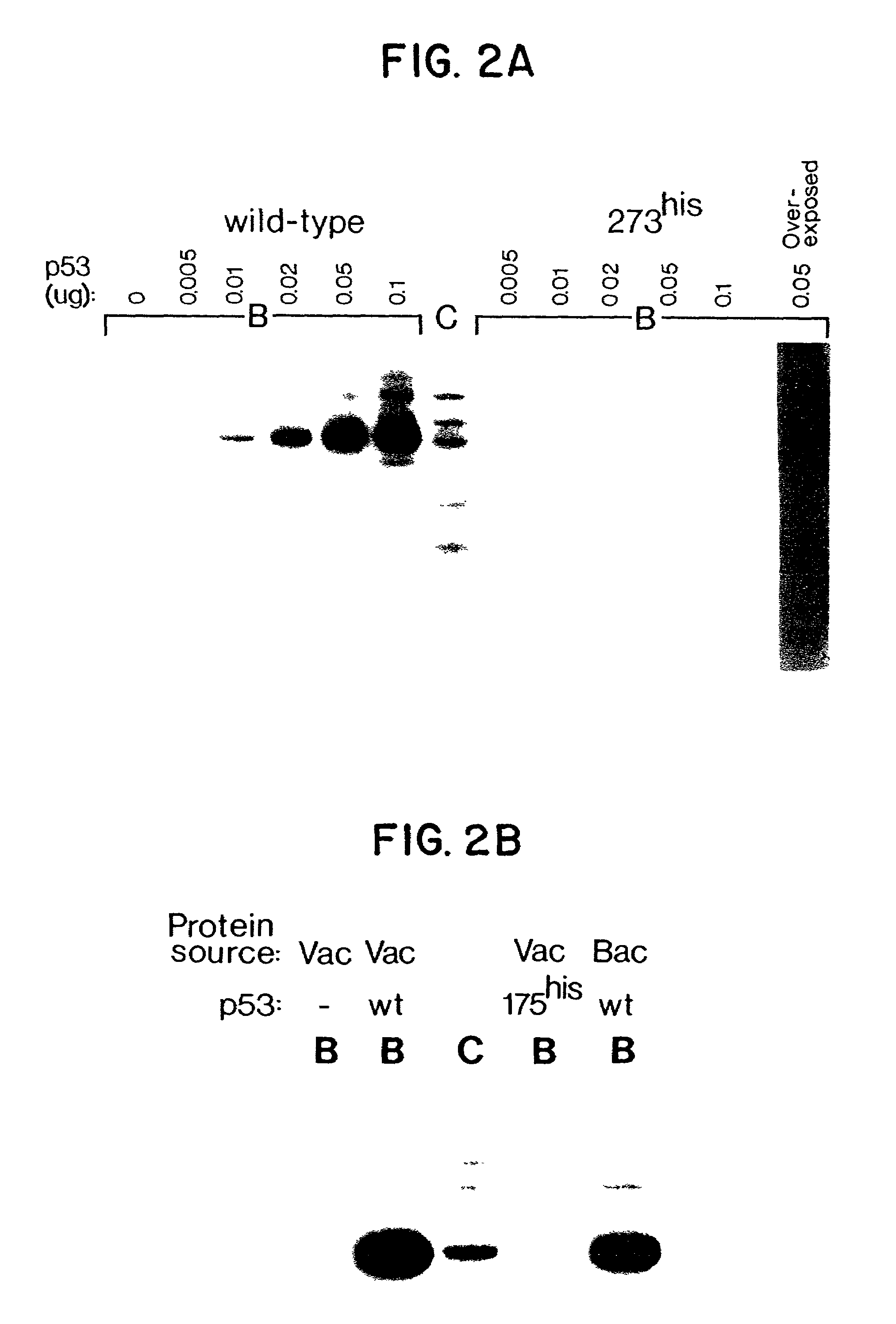
[0104]The example demonstrates that p53 mutant proteins found in human tumors fail to bind to fragment A.
[0105]Increasing quantities of wild-type and mutant 273his p53 protein, affinity purified from a baculovirus expression system, were used to immunoprecipitate labelled fragments from CBE. See FIG. 2A. The proportion of fragment A bound to wild-type p53 protein increased in tandem with the amount of p53 added to the assay mixture. (FIG. 2A) In contrast, fragment A did not specifically bind to a mutant form of p53 (273his) protein even at the highest p53 protein concentration used. The 273his mutation is the most common p53 mutant identified in human tumors. Another p53 mutant (175his) protein commonly found in human tumors also failed to bind to fragment A (FIG. 1B).
PUM
| Property | Measurement | Unit |
|---|---|---|
| Time | aaaaa | aaaaa |
| Time | aaaaa | aaaaa |
| Electric charge | aaaaa | aaaaa |
Abstract
Description
Claims
Application Information