
Methods and composition for restoring conformational stability of a protein of the p53 family

a technology of conformational stability and p53 family, which is applied in the field of cancer treatment, can solve the problems of less capable of retaining native conformation, uncontrolled proliferation of affected cells and tumor growth, and impaired cell function, and achieve negative dna binding effect, enhanced conformational stability, and wild-type activity.

Inactive Publication Date: 2002-04-25
RASTINEJAD FARZAN +3
View PDF28 Cites 34 Cited by
  • Summary
  • Abstract
  • Description
  • Claims
  • Application Information

AI Technical Summary

Benefits of technology

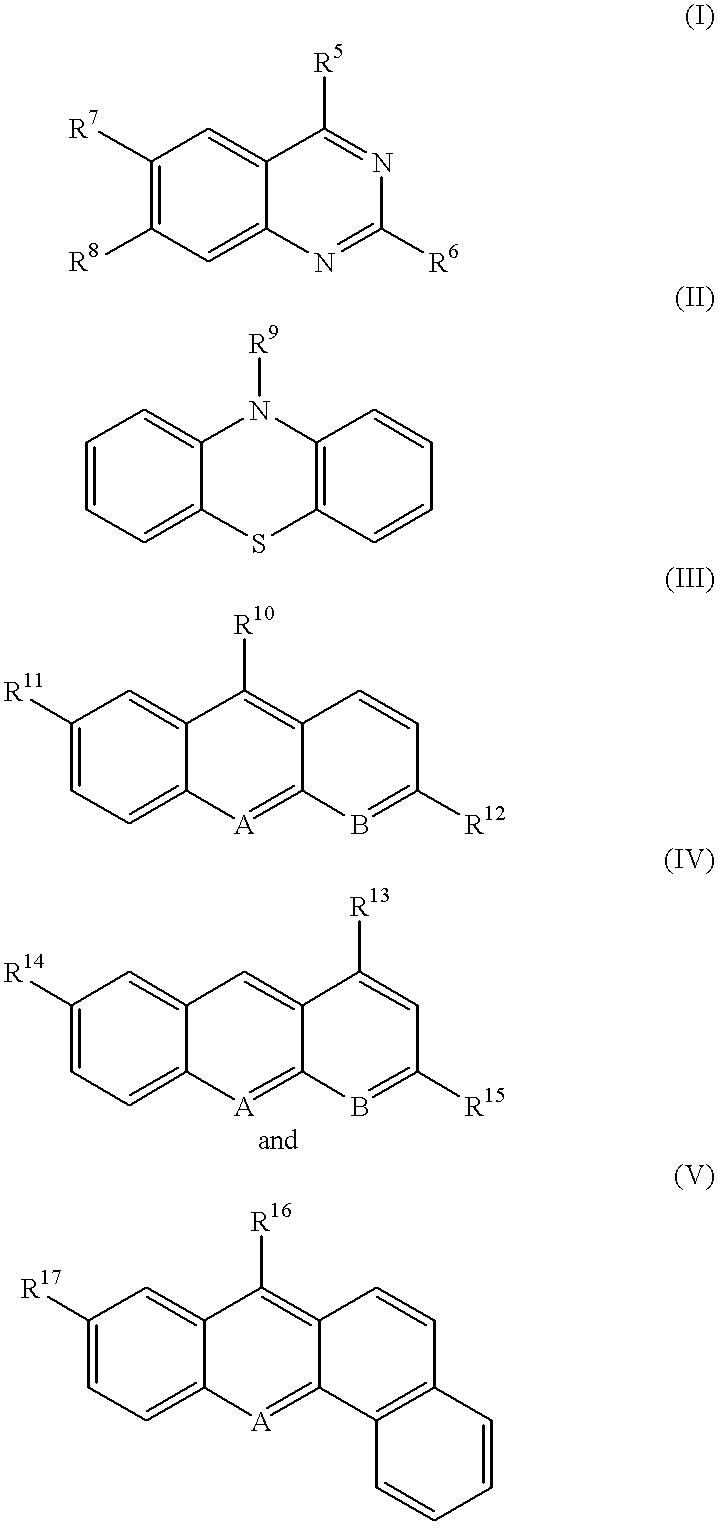
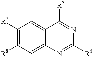
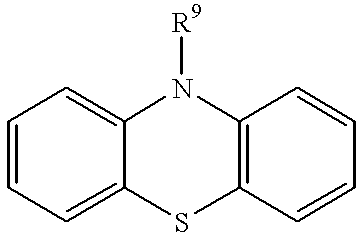
[0080] In yet another aspect, the invention includes the use of the compounds of the invention to inhibit tumor growth and / or treat cancer. A particular advantage of the invention is that the compounds so identified using the methods herein have been shown to stabilize the active conformation of not only wild-type p53 DBD and the mutant p53 DBD used in the screens, but also other mutant p53s and p53 DBDs. Therefore, the compounds so identified have broad applicability in treating varied cancers.
[0081] The present invention also provides a novel way of screening for compounds that promote the wild-type conformation of a protein of the p53 family and can restore wild-type activity to mutant proteins of the p53 family. Compounds identified using the methods of the invention are useful for treating diseases such as cancer that are associated with defects in activity of proteins of the p53 family.
[0082] The methods of the invention entail screening compounds for those that interact directly with a protein of the p53 family. Such methods can use a full length protein of the p53 family (mutant or wild-type) for screening purposes, or a deletion derivative containing at least the DBD and optionally the N terminal and / or C terminal domains. However, in a preferred aspect of the invention, the screens make use of a polypeptide fragment of a protein of the p53 family that contains only the DBD without the intact N or C terminal domains. Accordingly, for purposes of this Application, the term the DNA binding domain" or "the DBD" is understood to include just the DBD of a protein of the p53 family, without an intact N or C terminus (unless indicated otherwise). Such DBD domains may, however, be fused to heterologous polypeptides depending upon the assay format (e.g., a FLAG epitope or a glutathione-S-transferas-e protein). Additionally. rather than merely removing a negative regulatory effect on DNA binding, the methods and compounds of the invention promote enhanced conformational stability of both wild-type and mutant proteins of the p53 family.
[0083] Accordingly, in one aspect illustrated below by way of a non-limiting working example, the invention provides a method of screening for compounds that specifically interact with the p53 DBD, and measuring the conformation of the p53 DBD in the presence of the test compound. Optionally, the p53 DBD is a mutant p53 DBD. However. wild-type p53 DBD is easier to overproduce in large quantities. Although the screening assay can be performed in a cell-based format, for high-throughput screens specific to compounds that target the p53 DBD, an in vitro based assay is most direct and desired. Compounds identified in an initial screen against the p53 DBD can be further tested for their effects on the function of intact p53 (including p53 missense mutants). Compounds identified using these methods are also within the scope of the invention.
[0084] For purposes of the instant invention, assays for compounds that interact with the DNA binding domain of a protein of the p53 family are designed such that compounds uncovered are those that specifically target the DBD and not other domains of the protein. For example, a compound that specifically "interacts with" or "acts on" the DBD need not necessarily bind stably to the DBD (although it may); it is sufficient for the compound to have some effect on the conformation of a protein of the p53 family in the presence of the compound. Accordingly, compounds may be first screened for interaction with the DBD, and then assayed for their effect on conformation, or these two screening steps may be performed simultaneously by using a conformational change in the presence of the compound to also detect interaction with the DBD.
[0085] The term specific interaction in this application is used to exclude unspecific forms of binding including the type known to occur between hydrophobic compounds and proteins through nonselective hydrophobic interactions. The term specific interaction is further used to distinguish the properties of the compounds of this invention from compounds that affect protein thermostability by changing the chemical properties of the bulk solvent. Such molecules excluded from the scope of this aspect of the invention therefore include thermostabilizing agents such as glycerol, trimethylamine -oxide, and deuterated water. Compounds that specifically interact with a protein of the p53 family will show an effect at much lower concentrations than such bulk solvents or non-specific hydrophobic interactions. For example, glycerol is effective at 600 mM. However, effects of compounds that specifically interact with a protein of the p53 family will be observed at concentrations of the compound lower than 1 mM, preferably lower than 100 micomolar, and more preferably lower than 10 micromolar in in vitro or cell-based assays.

Problems solved by technology

For example, Alzheimer's disease is caused by misfolding and subsequent aggregation of beta-amyloid protein, leading to impairment of cell function.
Proteins that adopt abnormal conformations may do so either because they are inherently susceptible to misfolding or because they have mutations that thermodynamically destabilize the mutant protein relative to wild-type protein.
Loss of p53 activity can lead to uncontrolled proliferation of the affected cells and tumor growth.
Additionally, p53 mutants often associate with heat shock proteins in cells, leading to speculation that they are less capable of retaining native conformation (Finlay et al., 1988, Molecular and Cellular Biology 8:531-39).
Further, the discovery of such compounds has been precluded by the lack of a high through-put screen or assay.

Method used


Image

  • Methods and composition for restoring conformational stability of a protein of the p53 family
  • Methods and composition for restoring conformational stability of a protein of the p53 family
  • Methods and composition for restoring conformational stability of a protein of the p53 family

Examples


example 1

VI. EXAMPLE 1

p53 DBD Thermostabilization Assay

[0186] A high through-put assay using wild-type p53 DBD was developed. Pharmacological compounds were screened using the assay, and those compounds that stabilized the active conformation of the DBD were scored as hits.

[0187] A. Materials and Methods

[0188] Thermostabilization Assay. Recombinant DBD (residues 94-312) from wild-tvpe and mutant p53 proteins and FLAG-tagged p53 DBD were prepared as described (Pavletich et al., 1993, Genes and Dev. 7, 2556-2564; Bullock et al., 1997, supra.). Mutant proteins used were 143A, 173A, 175S. 249S, and 273H. A number of small molecule organic compounds were tested. Compound stocks were dissolved in DMSO at 10 mg / ml and diluted prior to use. The proteins (0.25-1.0 ng / well) were diluted in a buffer containing 25 mM HEPES, pH 6.8, 150 mM KCl, 10 mM dithiothreitol and attached in 50 ul to Reacti-Bind microtiter plates (Pierce) for 35 minutes on ice. The wells were rinsed with 25 mM HEPES, pH 6.8, 150 mM...

example 2

VII. EXAMPLE 2

Determination of p53 Conformation in Cells and Tumors

[0198] In this example and the examples that follow, prototype compounds are shown to function at low micromolar concentrations to modulate mutant p53 in living cells and in tumors and to suppress the growth of tumors with naturally mutated p53.

[0199] A. Materials and Methods

[0200] Cell Culture. All cell lines were obtained from the ATCC and grown in the recommended media with 10 percent fetal calf serum (Gibco BRL).

[0201] Determination of p53 Conforniation Approximately 1.times.10.sup.7H1299 / Reporter+Mutant p53 cells were treated overnight, rinsed three times with cold Tris buffered saline, and lysed in 1.5 ml of hypotonic lysis buffer (20 mM HEPES, pH 7.4, 10 mM NaCl, 20 percent glycerol, 0.2 mM EDTA, 0.1 percent Triton-X 100, 10 mM dithiothreitol with protease inhibitors). Cells were pelleted in microfuge tubes at 2000 rpm for 5 minutes at 4.degree. C. and nuclear extracts were prepared by resuspending the pellet...

example 3

VIII. EXAMPLE 3

Restoration of p53 Function

[0206] A. Materials and Methods

[0207] Transactivation assays. Cells were transfected with expression plasmids encoding mutant p53 proteins (173A, 249S) and a neomycin selectable marker using DOTAP cationic lipid transfection-reagent (Boehringer Mannheim) or calcium phosphate. Cells were also transfected with a plasmid encoding the hygromycin resistance marker and a p53 reporter gene comprised of four copies of a p53 binding sequence corresponding to a p53 binding sequence in the promoter region of the Herpes Simplex virus thymidine kinase gene (base numbers 26 to 58 of GenBank accession no. S57428 thymidine kinase, which begins with he sequence GCCTTGCCT and ends with the sequence TGCCTTTTC) placed upstream of he SV40 basal promoter driving the luciferase gene. A matched cell pair was prepared by transfecting a clone of cells with the reporter construct with an additional construct for mutant p53 expression. Transfected clones were selected ...


PUM

PropertyMeasurementUnit
lengthaaaaaaaaaa
lengthaaaaaaaaaa
lengthaaaaaaaaaa

Abstract

The invention is in the field of cancer treatment. In particular, the present invention provides pharmaceutical compounds capable of interacting with mutant and non-mutant forms of cancer-related regulatory proteins such that the mutant protein regains the capacitv to properly interact with other macromolecules thereby restoring or stabilizing all or a portion of its wild type activity. Regulatory proteins include members of the p53 protein family such as. for example, p53, p63 and p73. The compounds of the invention are useful for cancer treatment. Methods for screening for such pharmacological compounds are also provided.

Description

I. FIELD OF THE IVNENTION[0001] The invention is in the field of cancer treatment. The present invention provides organic non-peptide compounds capable of interacting with a tumor suppressor protein of the p53 family and stabilizing a functional conformation therein. The invention is particularly applicable to stabilizing mutant forms of tumor suppressor proteins in patients where correcting the functional capacity of such proteins can facilitate treatment for cancer. Methods for screening for such compounds are also provided.II. BACKGROUND OF THE INVENTION[0002] The primary structure of a protein is the particular sequence of amino acid building blocks that are linked together to form the protein's polypeptide chain(s). These polypetide chains are, in turn, folded into a three-dimensional structure. A number of diverse diseases are now thought to arise from a conformational perturbation in the three-dimensional structure of a cellular protein (see for reviews Thomas et al., 1995, T...

Claims


Application Information

Patent Timeline
Patent Type & Authority Applications(United States)
IPC IPC(8): A61K31/00A61K31/47A61K31/473A61K31/517A61K31/5415A61K45/08A61P3/06C12N15/09A61P7/04A61P21/04A61P25/00A61P25/28A61P27/02A61P27/12A61P35/00A61P43/00C07D219/12C07D221/08C07D221/18C07D239/94C07D279/26C07D417/12C07K14/47
CPCA61K31/473A61K31/5415A61K31/517A61P21/04A61P25/00A61P25/28A61P27/02A61P27/12A61P35/00A61P3/06A61P43/00A61P7/04
Inventor RASTINEJAD, FARZANFOSTER, BARBARA A.COFFEY, HEATHER A.CONNELL, RICHARD D.
Owner RASTINEJAD FARZAN
Learn More