
Color Doppler ultrasound imaging module and method

A technology of color Doppler and ultrasound imaging, applied in the direction of blood flow measurement devices, etc., can solve problems such as difficult to make diagnostic conclusions, time-consuming, and large number of parameters

Active Publication Date: 2012-07-04
CHISON MEDICAL TECH CO LTD
View PDF8 Cites 19 Cited by
  • Summary
  • Abstract
  • Description
  • Claims
  • Application Information

AI Technical Summary

Problems solved by technology

[0004] Although the color Doppler technology plays an important role in the diagnosis of the patient's blood flow or moving tissue, there are still the following deficiencies: First, in the color Doppler mode of the current ultrasonic diagnostic instrument, the doctor manually selects the corresponding Knob or button to determine whether to display color blood flow imaging, velocity variance imaging or energy imaging; secondly, in the color Doppler display, the displayed velocity range is determined by the pulse wave repetition frequency (PRF), while At the beginning of the examination, the speed range of the blood flow cannot be predicted accurately, and the doctor often sets a certain initial speed range based on experience, and then must be in the best state by continuously adjusting various parameter values ​​during the examination
In this way, on the one hand, the doctor has a large manual workload during the entire examination process. When there are a large number of patients diagnosed, it is inevitable that due to human factors, improper adjustment of the instrument may occur, so that some artifacts may appear during the application of color Doppler. , making it difficult to make correct diagnostic conclusions. On the other hand, there are many parameters that need to be adjusted manually, which is time-consuming and reduces the work efficiency of doctors to a certain extent.

Method used


Image

  • Color Doppler ultrasound imaging module and method
  • Color Doppler ultrasound imaging module and method
  • Color Doppler ultrasound imaging module and method

Examples


Embodiment Construction

[0041] Various details involved in the technical solution of the present invention will be described in detail below in conjunction with the drawings and embodiments.

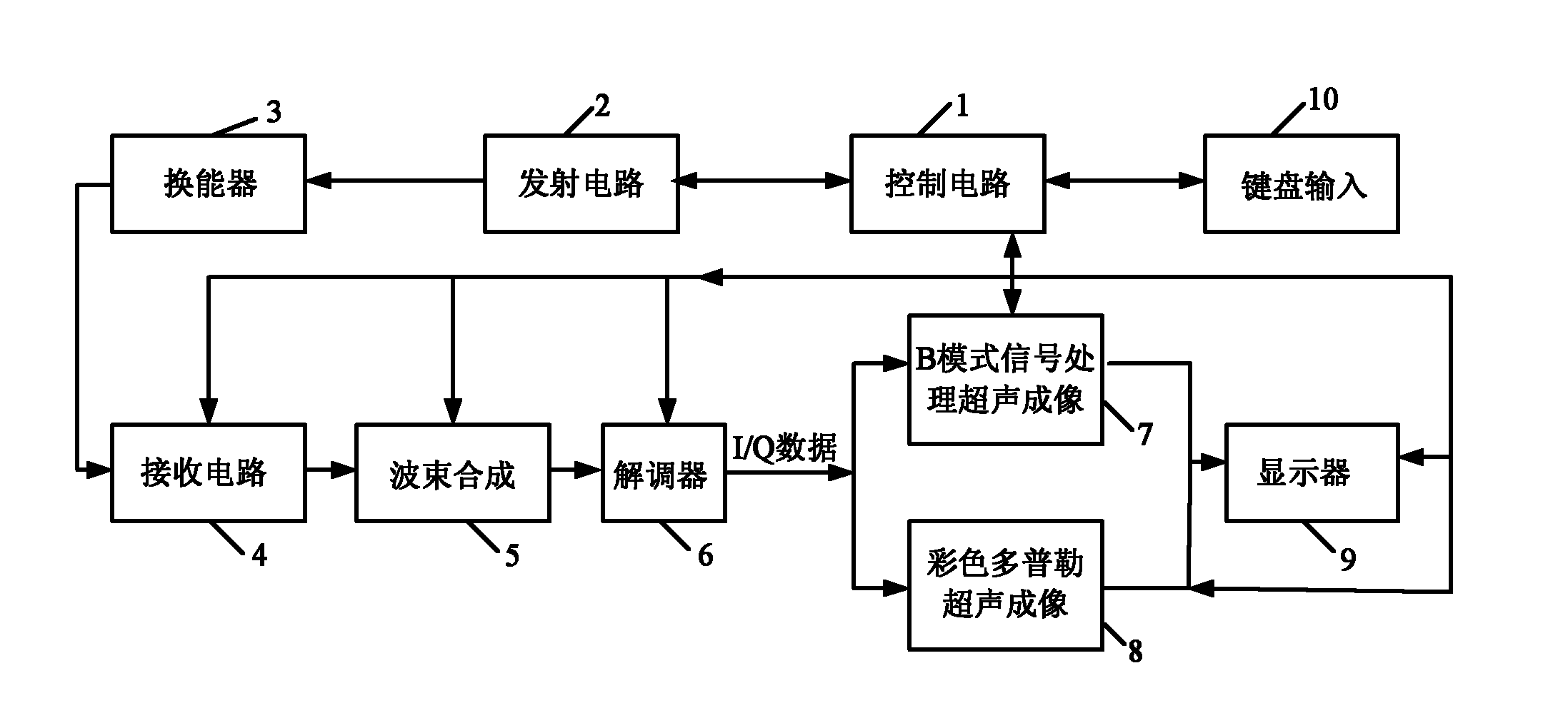
[0042] Such as figure 1 As shown, a system of ultrasonic equipment includes: control circuit 1, transmitting circuit 2, transducer 3, receiving circuit 4, beamforming 5, demodulator 6, B-mode signal processing ultrasonic imaging module 7, color Doppler Ultrasonic imaging module 8 , display 9 and keyboard input 10 . Transducer 3 (also called probe) is an ultrasonic transmitting and receiving device, which can convert electrical energy into acoustic energy, and can also convert acoustic energy into electrical energy. 3 Send an electrical signal, which is converted into ultrasonic waves by the transducer 3 and emitted, and the receiving circuit 4 is responsible for receiving the echo signal (which has been converted into an electrical signal by the transducer) from the transducer 3, and amplifies it, Digital-to-...


PUM


Abstract

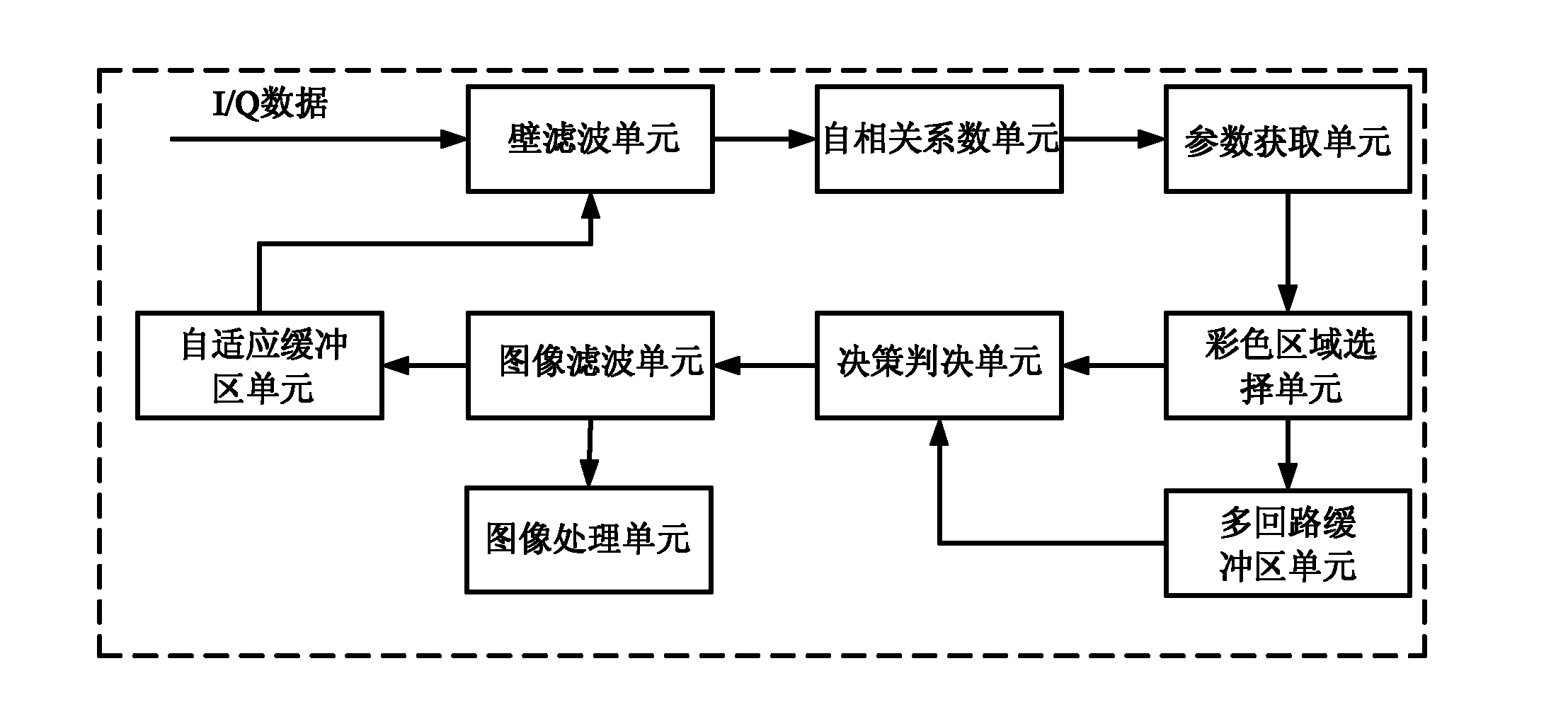
The invention discloses a color Doppler ultrasound imaging module and method. The module comprises a wall filtering unit, an auto-correlation coefficient unit, a parameter acquisition unit, a color region selection unit, a decision-making and judging unit, a multi-loop buffer region unit, an image filtering unit, a self-adapting buffer region unit and an image processing unit, wherein the wall filtering unit, the auto-correlation coefficient unit, the parameter acquisition unit and the color region selection unit are connected in sequence; the color region selection unit is connected with the decision-making and judging unit and the multi-loop buffer region unit respectively; the output end of the multi-loop buffer region unit is connected with the input end of the decision-making and judging unit; the output end of the decision-making and judging unit is connected with the image filtering unit; the output end of the image filtering unit is connected with the self-adapting buffer region unit and the image processing unit respectively; and the output end of the self-adapting buffer region unit is connected with the wall filtering unit. The invention has the advantages that the color Doppler ultrasound imaging effect is improved and the implementation of color Doppler ultrasound imaging is easier.

Description

technical field [0001] The invention relates to an ultrasonic imaging module and method, in particular to a color Doppler ultrasonic imaging module and method. Background technique [0002] Color Doppler is the use of autocorrelation technology for Doppler signal processing. The average velocity, variance and energy of blood flow obtained by autocorrelation technology are color-coded and superimposed on the two-dimensional grayscale image in real time to form a color Doppler image. Ultrasound blood flow images. [0003] In a color Doppler image, blood flow toward the probe is shown as a red signal, and blood flow away from the probe is shown as a blue signal. In addition to the above information, color Doppler images can also display information such as the speed of blood flow and reflect the nature of blood flow. [0004] Although the color Doppler technology plays an important role in the diagnosis of the patient's blood flow or moving tissue, there are still the followi...

Claims


Application Information

Patent Timeline
IPC IPC(8): A61B8/06
Inventor 赵丹华张勇赵明昌陆坚
Owner CHISON MEDICAL TECH CO LTD
Learn More