Ultrasonic contrast video analysis method and system

A technology of contrast-enhanced ultrasound and analysis methods, which is applied in image analysis, ultrasound/sound wave/infrasonic diagnosis, sound wave diagnosis, etc., to achieve the effect of improving diagnostic accuracy

Inactive Publication Date: 2012-07-11
CHONGQING AMBITION TECH
View PDF4 Cites 9 Cited by
  • Summary
  • Abstract
  • Description
  • Claims
  • Application Information

AI Technical Summary

Problems solved by technology

However, it is not yet possible to realize the off-line processing and analysis of ultrasound-enhanced video files

Method used


Image

  • Ultrasonic contrast video analysis method and system
  • Ultrasonic contrast video analysis method and system
  • Ultrasonic contrast video analysis method and system

Examples


Embodiment Construction

[0026] Preferred embodiments of the present invention will be described in detail below.

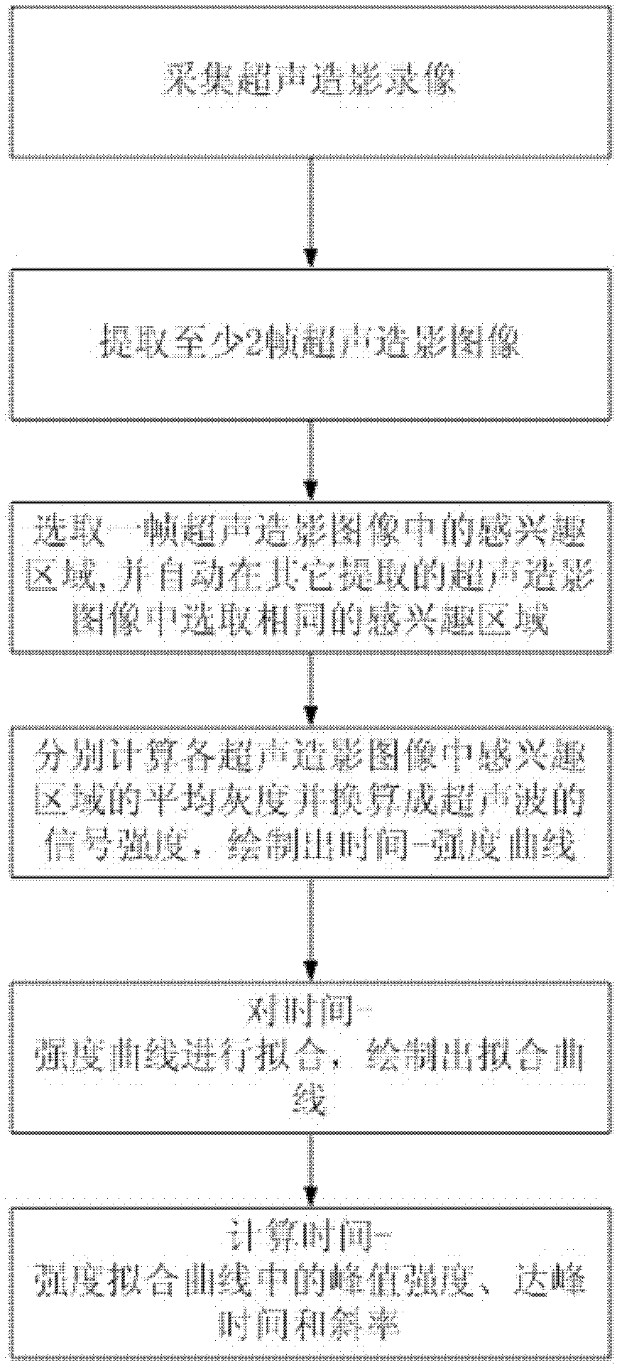
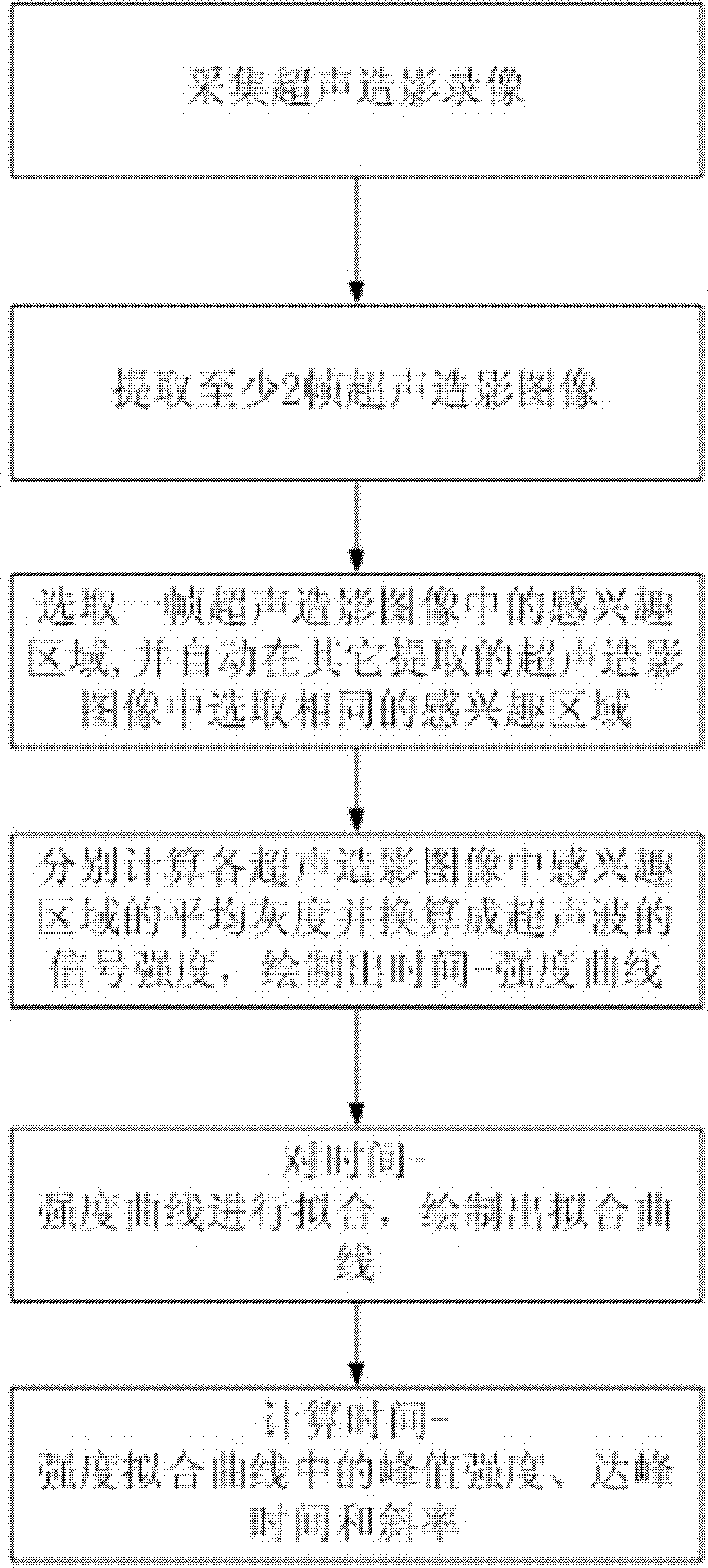
[0027] see figure 1 , the ultrasonic contrast-enhanced video analysis method of the present embodiment includes the following steps:

[0028] 1) Acquisition of contrast-enhanced ultrasound video;

[0029] 2) From the ultrasound-contrast-enhanced video collected in step 1), extract each frame of ultrasound-enhanced images in chronological order;

[0030] 3) According to the position, shape and size of the region of interest (ROI) selected by the operator in the first frame of image, automatically select the region of interest with the same position, shape and size from each frame of contrast-enhanced ultrasound image;

[0031] 4) Calculate the average gray level of the region of interest in each contrast-enhanced ultrasound image and convert it into the signal strength (dB) of the ultrasound through the ultrasound dynamic range, and draw the time-intensity curve;

[0032] 5) Curve fitt...


PUM


Abstract

The invention relates to the technical field of ultrasonic contrast, in particular to an ultrasonic contrast video analysis method and system, which can quantitatively analyze ultrasonic contrast video. The method comprises collecting ultrasonic contrast video; extracting at least two frames of ultrasonic contrast images from the collected ultrasonic contrast video according to time sequence; selecting an area of interest in one frame of ultrasonic contrast image, and automatically selecting the same area of interest in the other frames of ultrasonic contrast images; calculating average grayscales of the areas of interest in the ultrasonic contrast images, respectively, converting to ultrasonic signal intensity, and working out a time-intensity curve; fitting the time-intensity curve, and working out a fitted curve; and calculating relevant parameters in the fitted curve. The system comprises a video collection module, an image extraction module, an area of interest selection module, an intensity analysis module, a time-intensity curve plotting module, a curve fitting module and a curve analysis module.

Description

technical field [0001] The invention relates to the technical field of contrast-enhanced ultrasound, in particular to a video analysis method and system for contrast-enhanced ultrasound. Background technique [0002] The "contrast acoustic imaging" technology has been used by clinicians in the United States as one of the routine testing items. This technique has the following advantages: At present, conventional ultrasonography cannot detect the blood flow with very low velocity and flow in the tissue, such as the blood perfusion of capillaries in the tissue. Contrast-enhanced ultrasound can display the blood perfusion characteristics of diseased tissue, increase the contrast difference between diseased tissue and normal tissue, thereby improving the ability of ultrasound to detect lesions and make qualitative diagnosis. The accuracy of evaluation of local treatment effect is comparable to that of enhanced CT and enhanced MR, and it can also play a very good auxiliary role ...

Claims


Application Information

Patent Timeline
Patent Type & Authority Applications(China)
IPC IPC(8): A61B8/00G06T7/00
Inventor 郑元义何发东
Owner CHONGQING AMBITION TECH
Why Eureka
Try Eureka