Segmentation method of viscera and internal blood vessels thereof in surgical planning system

A surgical planning, visceral technology, applied in the field of medical image processing, can solve difficult and time-consuming problems

Active Publication Date: 2012-09-12
SUZHOU DIKAIER MEDICAL TECH
View PDF3 Cites 21 Cited by
  • Summary
  • Abstract
  • Description
  • Claims
  • Application Information

AI Technical Summary

Problems solved by technology

[0008] The emergence of semi-supervised algorithms is mainly based on the following considerations: In most cases, in order to achieve better results for fully supervised classification algorithms, a

Method used


Image

  • Segmentation method of viscera and internal blood vessels thereof in surgical planning system
  • Segmentation method of viscera and internal blood vessels thereof in surgical planning system
  • Segmentation method of viscera and internal blood vessels thereof in surgical planning system

Examples


Embodiment Construction

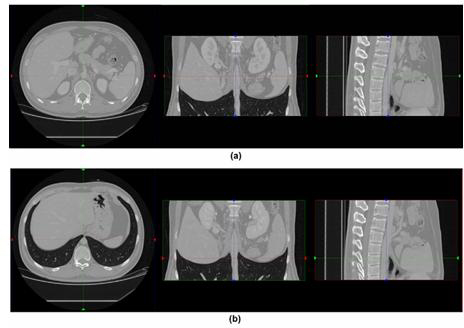
[0046] This embodiment is realized in a computer whose CPU is Intel(R) Core?? i3-2100 3.10GHz and internal memory is 2.0GB, the programming language is C++, and the three-dimensional visceral CT image to be segmented, such as figure 2 The original abdominal CT image of a patient to be segmented is as follows:

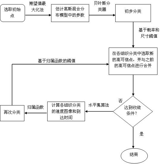
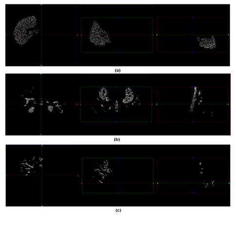
[0047] A segmentation method of viscera and its internal blood vessels in the surgical planning system, which is carried out directly on the running computer. First, the three-phase image of the patient's original CT scan is read into the system, and the three-dimensional visceral CT image and its internal organs are based on the minimum supervised classification. Tubular tissue, formulate the image segmentation method of liver parenchyma, portal vein and hepatic vein, which applies statistical and spatial information methods, introduces high-confidence points and gray-scale-based fast-moving arrival time, and obtains segmentation results, according to the following ste...


PUM


Abstract

The invention relates to a segmentation method of medical images, and specifically relates to a segmentation method of viscera and internal blood vessels thereof in a surgical planning system. The method designs a segmentation method of images of liver parenchyma, portal veins and hepatic veins based on minimal supervised classification of the three-dimensional CT images of the viscera and internal tubular tissues thereof. The method applies methods of statistics and spatial information, introduces high credible points, and obtains segmentation results based on arrival time of fast march of grey level. The method assumes that spatially adjacent points belong to a same organ, and performs segmentation calculation on the images by using a classification algorithm based on the above assumption, wherein the segmentation calculation comprises the steps of: acquiring estimated values of parameters of a Gaussian mixture distribution model; selecting the high credible points; and calculating the mean value of the grey level of the images and calculating the arrival time by using a fast marching algorithm to obtain arrival time images of three tissue classifications. According to the invention, good robustness and good anti-noise interference capability are obtained, accumulated errors are eliminated effectively, classification accuracy is improved, and great application value in clinical treatment is achieved.

Description

technical field [0001] The segmentation method of viscera and its internal blood vessels in the surgical planning system of the present invention relates to a medical image segmentation method, which is used in the surgical planning system, especially to segment the liver parenchyma, hepatic portal vein and hepatic A vein belongs to the technical field of medical image processing. Background technique [0002] The important work of computer-aided liver surgery planning is to accurately segment the tree structure of liver parenchyma, hepatic portal vein and hepatic vein from original medical images, and make the extracted results as close as possible to the real anatomical structure. In the segmentation problem of the hepatic portal vein and hepatic vein, because the spatial structure and distribution of the hepatic portal vein and hepatic vein vary greatly among different individuals, the gray distribution of the hepatic portal vein and hepatic vein is close to the liver par...

Claims


Application Information

Patent Timeline
IPC IPC(8): G06K9/62G06T7/00
Inventor 顾力栩
Owner SUZHOU DIKAIER MEDICAL TECH
Why Eureka
Try Eureka