System and method for four dimensional angiography and fluoroscopy

一种血管、造影剂的技术,应用在用于放射诊断的仪器、应用、心导管等方向,能够解决血栓栓塞并发症风险升高等问题

Active Publication Date: 2012-09-26
MISTRETTA MEDICAL +1
View PDF2 Cites 13 Cited by
  • Summary
  • Abstract
  • Description
  • Claims
  • Application Information

AI Technical Summary

Problems solved by technology

Not only does this use a large amount of contrast media, but the risk of thromboembolic complications increases with each injection

Method used


Image

  • System and method for four dimensional angiography and fluoroscopy
  • System and method for four dimensional angiography and fluoroscopy
  • System and method for four dimensional angiography and fluoroscopy

Examples


Embodiment Construction

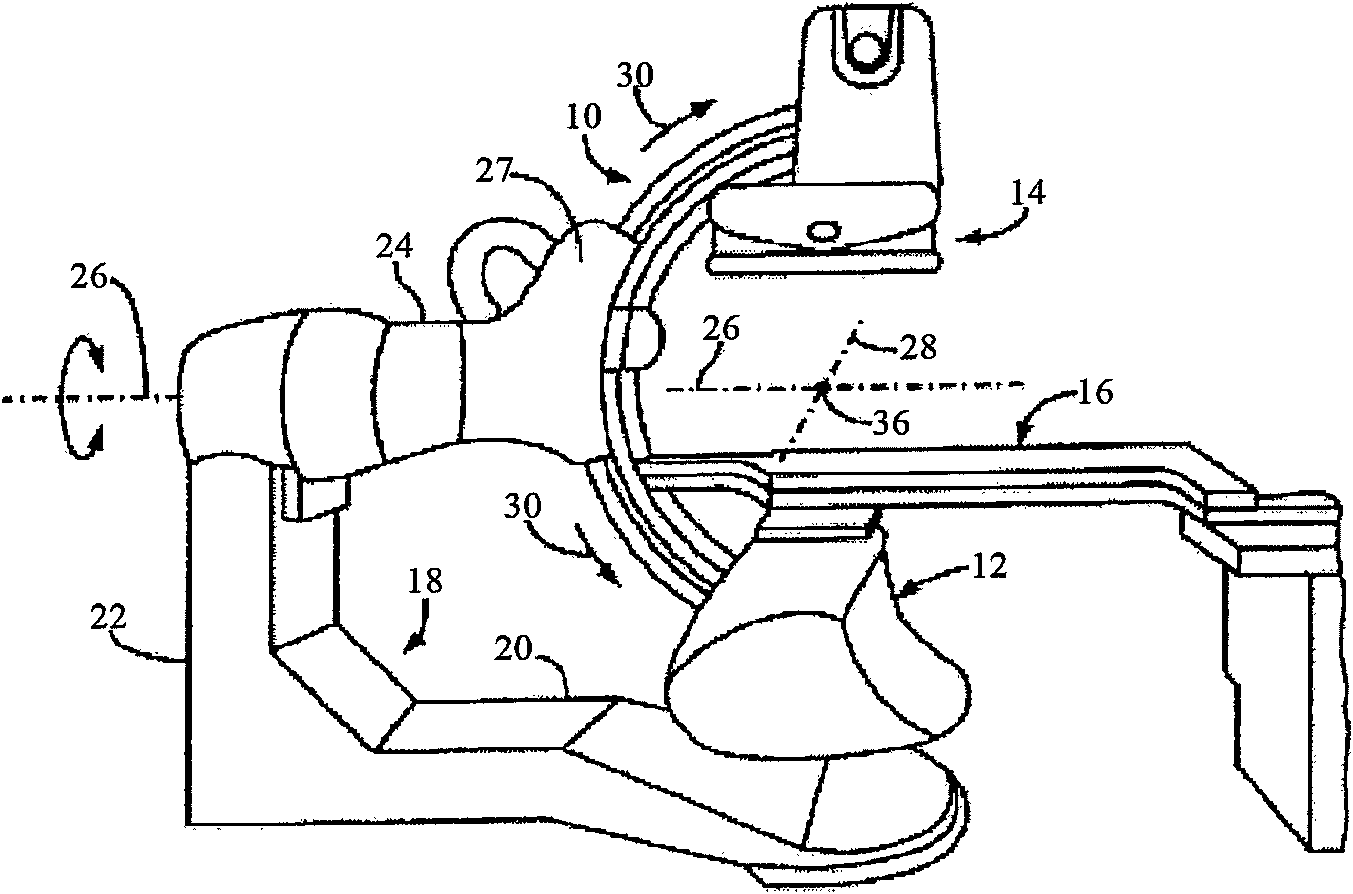
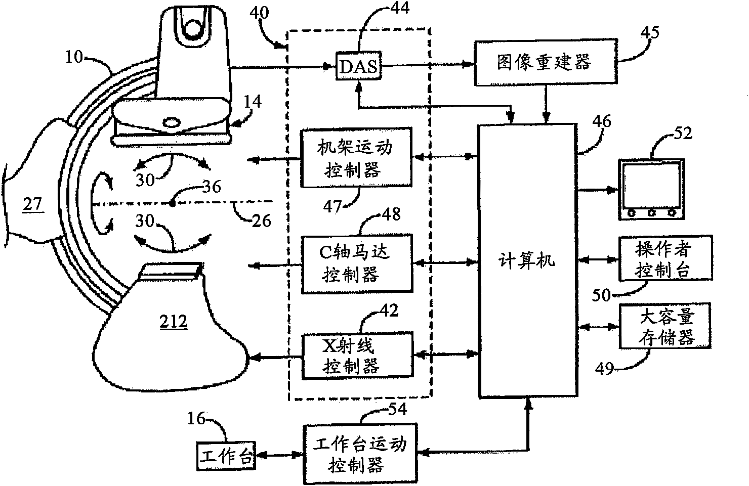
[0099] refer to Figure 1A , the present invention may employ a rotating X-ray system specifically designed for use with interventional procedures. It features a gantry having a C-arm 10 with an X-ray source assembly 12 at one end thereof and an X-ray detector array assembly 14 at its other end. The gantry enables the x-ray source 12 and detector 14 to be oriented in different positions and angles around the patient positioned on the table 16, while allowing the physician close access to the patient.

[0100]The frame includes an L-shaped base frame 18 having horizontal legs 20 extending below the table 16 and vertical legs extending upward at the ends of the horizontal legs 20 spaced from the table 16. twenty two. A support arm 24 is rotatably secured to the upper end of the vertical leg 22 for rotation about a horizontal pivot axis 26 .

[0101] Pivot axis 26 is aligned with the centerline of table 16 and arm 24 extends radially outward from pivot axis 26 to support a C-a...


PUM


Abstract

A method for generating time-resolved 3D medical images of a subject by imparting temporal information from a time-series of 2D medical images into 3D images of the subject. Generally speaking, this is achieved by acquiring image data using a medical imaging system, generating a time-series of 2D images of a ROI from at least a portion of the acquired image data, reconstructing a 3D image substantially without temporal resolution from the acquired image data, and selectively combining the time series of 2D images with the 3D image. Selective combination typically involves registering frames of the time-series of 2D images with the 3D image, projecting pixel values from the 2D image frames 'into' the 3D image, and weighting the 3D image with the projected pixel values for each frame of the time-series of 2D images. This method is particularly useful for generating 4D-DSA images (that is, time-resolved 3D-DSA images) from a time-series of 2D-DSA images acquired via single plane or biplane x-ray acquisitions with 3D images acquired via a rotational DSA acquisition. 4D-DSA images can be generated either by using multiple injections or by using a single injection by combining a time-series of 2D-DSA images generated from individual projections from a rotational x-ray acquisition with a 3D image reconstructed from substantially all of the projection views acquired during the rotational x-ray acquisition. These DSA images may have a spatial resolution on the order of 5123 pixels and a temporal resolution of about 30 frames per second, which represents an increase over traditional 3D-DSA frame rates by a factor of between 150 and 600.

Description

[0001] Cross References to Related Applications [0002] This application claims priority to U.S. Patent Application No. 12 / 692,340, filed January 22, 2010, entitled "SYSTEMAND METHOD FOR FOUR DIMENSIONAL ANGIOGRAPHY AND FLUOROSCOPY," which was filed August 17, 2009 Continuation-in-Part of US Patent Application Serial No. 12 / 542,376, entitled "SYSTEM AND METHOD OF TIME-RESOLVED, THREE-DIMENSIONAL ANGIOGRAPHY," each of which is hereby incorporated by reference in its entirety. The entire contents of US Patent Application Serial No. 12 / 542,376 are contained in Appendix I of the current file. [0003] The following descriptions are provided to help readers understand. None of the information presented or references cited is admitted to be prior art. technical field [0004] The present invention relates to angiography, and more particularly to systems and methods for generating time-resolved, three-dimensional (thus, forming four-dimensional) angiographic and fluoroscopic imag...

Claims


Application Information

Patent Timeline
Patent Type & Authority Applications(China)
IPC IPC(8): G06T15/00A61B6/00
CPCA61B6/486G06T11/008A61B6/5247A61B6/504A61B6/5223A61B6/487A61B6/4441Y10S378/901G06T19/00A61B6/481G06T2211/436G06T13/20A61M5/007G06T2211/404G06T2211/412A61B6/12A61B6/485A61B6/02A61B6/022
Inventor 查尔斯·A·米斯特雷塔查尔斯·M·斯特罗瑟
Owner MISTRETTA MEDICAL
Why Eureka
Try Eureka