Method, image processing device and computed tomography system for determining a proportion of necrotic tissue

A technique for necrotic tissue and tissue, applied in the field of computed tomography systems, to achieve the effects of good quality, avoiding motion artifacts, and fewer errors

Active Publication Date: 2015-06-17
SIEMENS HEALTHCARE GMBH
View PDF3 Cites 0 Cited by
  • Summary
  • Abstract
  • Description
  • Claims
  • Application Information

AI Technical Summary

Problems solved by technology

However, such methods require high computing power and unfortunately do not always provide flawless results.
Therefore, this method is rarely used in clinical practice

Method used


Image

  • Method, image processing device and computed tomography system for determining a proportion of necrotic tissue
  • Method, image processing device and computed tomography system for determining a proportion of necrotic tissue
  • Method, image processing device and computed tomography system for determining a proportion of necrotic tissue

Examples


Embodiment Construction

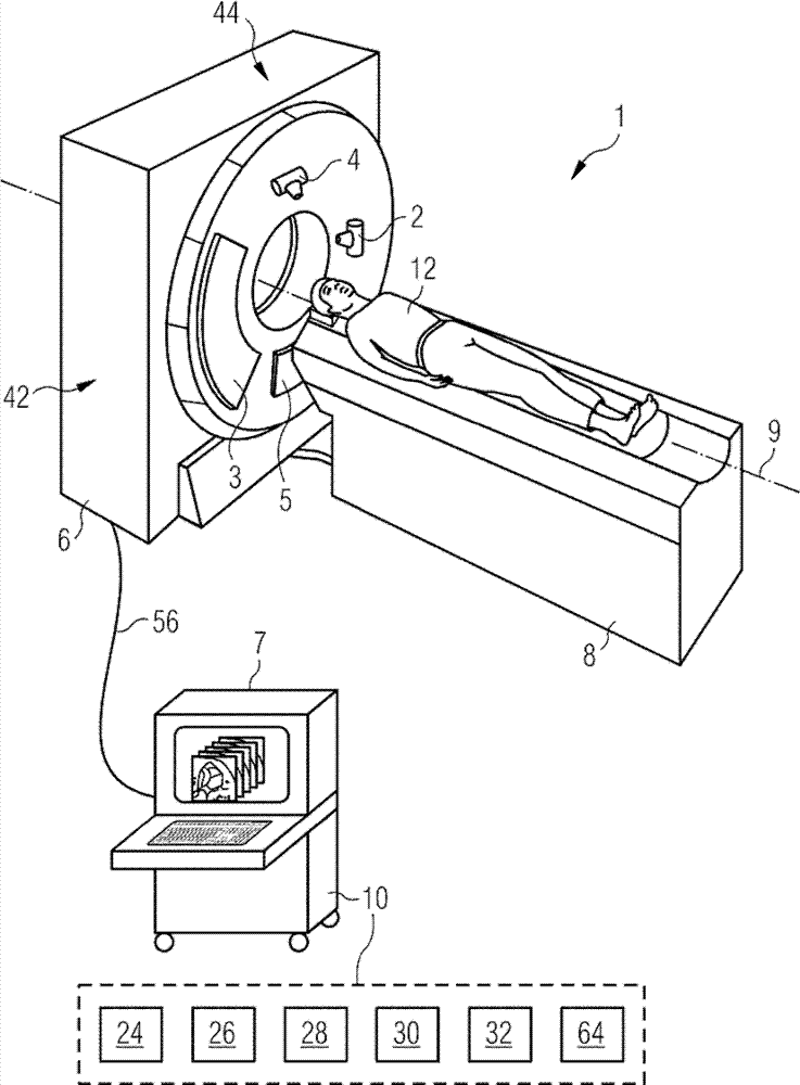
[0050] figure 1 The X-ray system shown in is a dual-source computed tomography machine 1 . It has a gantry (not exactly shown) mounted in a gantry housing 6 rotatable about a system axis 9 , on which two radiator / detector systems 42 , 44 are mounted angularly offset, which They are respectively formed by X-ray tubes 2, 4 and detectors 3, 5 arranged oppositely on the frame. The examination object 12 , here the patient, is located on a patient couch 8 which is movable along the system axis 9 and can be moved on this patient couch during the examination during the examination in the region of the radiation / detector systems 42 , 44 . field.

[0051] The control and possibly also the image reconstruction of the dual-source computed tomography apparatus 1 can be carried out by a conventional control device 7 which is specially equipped for the determination of necrotic tissue fractions for carrying out the method according to the invention. For this purpose, the control device 7 ad...


PUM


Abstract

An image processing device and method are disclosed for determining a proportion of necrotic tissue in a defined tissue area of an object under examination based on a high-energy image dataset and a low-energy image dataset, each recorded by way of x-ray measurements with different x-ray energies after a contrast medium has been applied to the object under examination. In at least one embodiment of the method, a virtual contrast medium image is determined from the high-energy image dataset and the low-energy image dataset and a segmentation image dataset is created, by the area of tissue being segmented. The segmentation result is transferred into the virtual contrast medium image for segmenting the tissue area in the virtual contrast medium image. Finally an analysis of values of the pixels lying in the segmented area is undertaken for identifying pixels which are to be assigned to necrotic tissue.

Description

technical field [0001] The invention relates to a method, an image processing device and a computed tomography system for determining necrotic tissue fractions in defined tissue regions of an examination object, in particular within tumors. Furthermore, the invention relates to a computer program product comprising program code segments for determining necrotic tissue fractions within a defined tissue region of an examination object, in particular within a tumor. Background technique [0002] In the treatment of tumor diseases, it is important to monitor the results of treatment through regular progress checks, so as to make timely judgments on whether to continue treatment or to change the treatment plan. But in addition to determining the size of the tumor tissue when assessing the outcome of treatment, it is also important to determine the necrotic tissue fraction of the tumor volume, since tumors respond differently to different forms of treatment and are not forced to c...

Claims


Application Information

Patent Timeline
Patent Type & Authority Patents(China)
IPC IPC(8): G06T7/00A61B6/03
CPCA61B6/12A61B6/482A61B6/5217G06T2207/10116G06T2207/10152G06T2207/30096G06T7/11G06T7/174G06T7/62G06T7/136A61B6/481G16H50/30
Inventor T.弗洛尔B.施米特M.塞德尔迈尔M.瓦格纳
Owner SIEMENS HEALTHCARE GMBH
Why Eureka
Try Eureka