Static CT (computed tomography) scanner and scattering X-photon correction method thereof
A scanner and static technology, applied in the field of medical equipment, can solve problems such as poor CT image quality, and achieve the effects of easy maintenance, reduced impact, and low operating costs
- Summary
- Abstract
- Description
- Claims
- Application Information
AI Technical Summary
Problems solved by technology
Method used
Image

Examples
Embodiment Construction
[0030] In order to make the object, technical solution and advantages of the present invention clearer, the present invention will be further described in detail below in conjunction with the accompanying drawings and embodiments. It should be understood that the specific embodiments described here are only used to explain the present invention, not to limit the present invention.
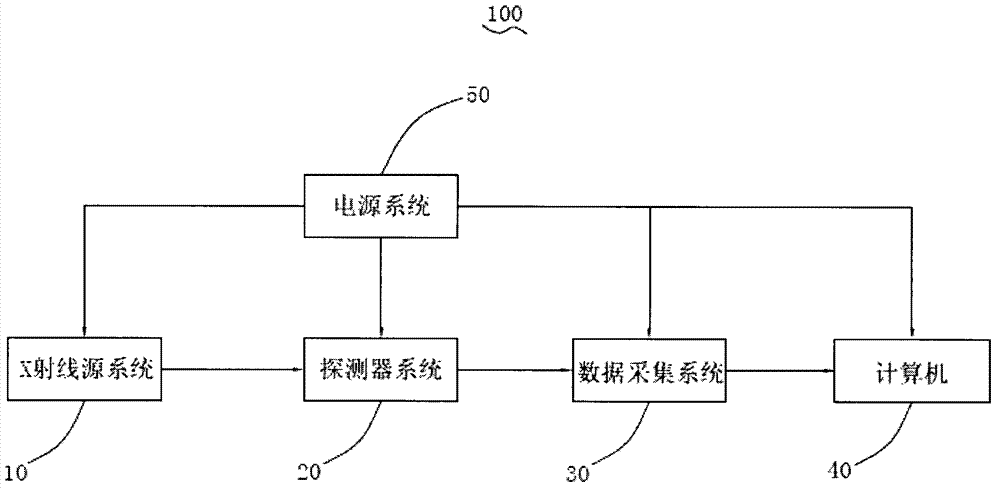
[0031] see image 3 , the first embodiment of the present invention provides a static CT scanner 100 , which includes an X-ray source system 10 , a detector system 20 , a data acquisition system 30 , a computer 40 and a power supply system 50 .
[0032] Please also refer to Figure 4 to Figure 6, the X-ray source system 10 includes an annular X-ray source 11 and an annular front collimator 13, the annular X-ray source 11 is used to emit X-rays, and it includes several X-ray source modules based on carbon nanotubes 111; the annular front collimator 13 is arranged at the exit of the annular X-ray s...
PUM
| Property | Measurement | Unit |
|---|---|---|
| Diameter range | aaaaa | aaaaa |
Abstract
Description
Claims
Application Information