Stereotaxic radiation therapy device for the head

A radiotherapy and stereotaxic technology, applied in X-ray/γ-ray/particle irradiation therapy, computed tomography scanner, echo tomography, etc., can solve the problems of dose error, insufficient head positioning accuracy, etc.

Active Publication Date: 2016-05-18
SUZHOU LINATECH MEDICAL SCI & TECH CO LTD
View PDF7 Cites 0 Cited by
  • Summary
  • Abstract
  • Description
  • Claims
  • Application Information

AI Technical Summary

Problems solved by technology

The disadvantage is: still due to the influence of the time axis in 4D space, the position of the tumor in the respiratory movement will change to a certain extent. If the method of 3D space is used for verification, the dose will have a certain error
[0006] To sum up, currently there is no stereotactic radiotherapy device specially designed for treating the head in the radiotherapy market. Generally, the head and body are integrated, and the positioning accuracy of the head is not high enough.

Method used


Image

  • Stereotaxic radiation therapy device for the head

Examples


Embodiment 1

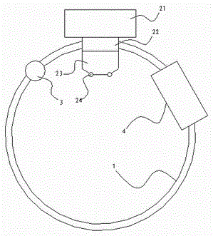
[0041] like figure 1 As shown, a head stereotaxic radiation therapy device includes:

[0042] A circular guide rail 1, an adapter 22 arranged on the circular guide rail, an accelerator 21 installed on the adapter, a digital image detection flat panel 4 mounted on the circular guide rail and sliding along it, and a digital image detection flat panel 4 installed on the circular guide rail and a ray emitting device 3 that can slide along it. The ray emitting device 3 is an X-ray emitting device.

[0043] A grating 23 is installed below the adapter 22 to control the area and shape of the rays passing through. The grating 23 is an electric multi-leaf grating. Two cameras 24 are also arranged on the grating 23 .

[0044] The adapter 22 is fixedly connected or integrated with the ring guide rail 1, and is directly installed on the head of the accelerator 21; the electric multi-leaf grating is directly installed on the adapter 22, and rotates with the ring guide rail 1; the camera...

Embodiment 2

[0060] The rest are the same as in Embodiment 1, the difference is that there are two digital image detection flat panels 4, one corresponds to the position of the accelerator 21 and is used in conjunction with the accelerator, and the other corresponds to the position of the ray emitting device 3 and is rotated synchronously and used in conjunction with it to improve The responsiveness of the entire device.

[0061] In the above embodiments, one, two, three or other numbers of the digital image detection panel 4, the ray emitting device 3, and the camera 24 can be determined as required.

[0062] The beneficial effect of adopting this technical solution is: the accelerator is integrated with the X-ray emitter and the digital image detection panel, and the three-dimensional image of the patient can be quickly obtained through the CBCT function. The tumor coordinates are quickly positioned on the isocenter; due to the unique structure of this technical solution, the CBCT only n...


PUM


Abstract

The invention discloses a radiotherapy device for stereo positioning of a head. An X-ray transmitter and a digital image detection panel are integrated on an accelerator, a three-dimensional image of a patient can be quickly obtained through a cone beam computed tomography (CBCT) function, and after the three-dimensional image is obtained, software quickly locates tumor coordinates in a data set on isocenters through an algorithm; and because the technical scheme is unique in structure, the three-dimensional image of the patient can be quickly obtained through only rotating the CBCT for 120 degrees, the consumed time is short, so a plurality of groups of three-dimensional images of the patient can be obtained during a time period, tumor position variations which are caused by breathing movement can be recorded in the plurality of groups of images, the tumor positions (registration) of the plurality of three-dimensional images are compared through the algorithm, and a four-dimensional plan is designed in a radio therapy planning system according to the displacement differences of the images groups.

Description

technical field [0001] The invention relates to a medical device, in particular to a head stereotaxic radiation therapy device used in conjunction with an accelerator therapy instrument. Background technique [0002] When performing radiation therapy on tumors (target areas), there are the following important indicators: [0003] First, the precise location of the tumor. In the 3D space, the doctor or physicist first scans the patient's CT images in the CT room, and reconstructs a series of CT images into a 3D image through an algorithm. At this time, the 3D coordinates of the tumor in the 3D image can be located. Then the doctor or physicist places the patient in the accelerator room for reset, and precisely coincides the three-dimensional coordinates of the tumor with the treatment isocenter of the accelerator. Finally start treatment. But in this method, there is a problem. When the patient breathes, the position of the tumor will change within a certain range with the...

Claims


Application Information

Patent Timeline
Patent Type & Authority Patents(China)
IPC IPC(8): A61N5/10A61B6/03
Inventor 姚毅
Owner SUZHOU LINATECH MEDICAL SCI & TECH CO LTD
Why Eureka
Try Eureka