
Full-automatic scanning system and method for microscopic section

A microsection and scanning system technology, applied in microscopes, optics, instruments, etc., can solve the problem of low speed, achieve the effect of reducing motion blur, fast image, and improving scanning speed

Active Publication Date: 2013-01-02
MOTIC CHINA GRP CO LTD
View PDF6 Cites 55 Cited by
  • Summary
  • Abstract
  • Description
  • Claims
  • Application Information

AI Technical Summary

Problems solved by technology

The disadvantage is: when shooting images at each new position, the stage needs to accelerate and then decelerate to the target position before stopping. This movement process takes about 100 milliseconds, and then it needs to wait for the system to completely stabilize for about 50 milliseconds
This stop-and-go, acceleration and deceleration movement mode and the resulting system vibration make the area scan digital slicing method slow, and it usually takes hundreds of seconds (several minutes) to complete the digital scanning process of a slice

Method used

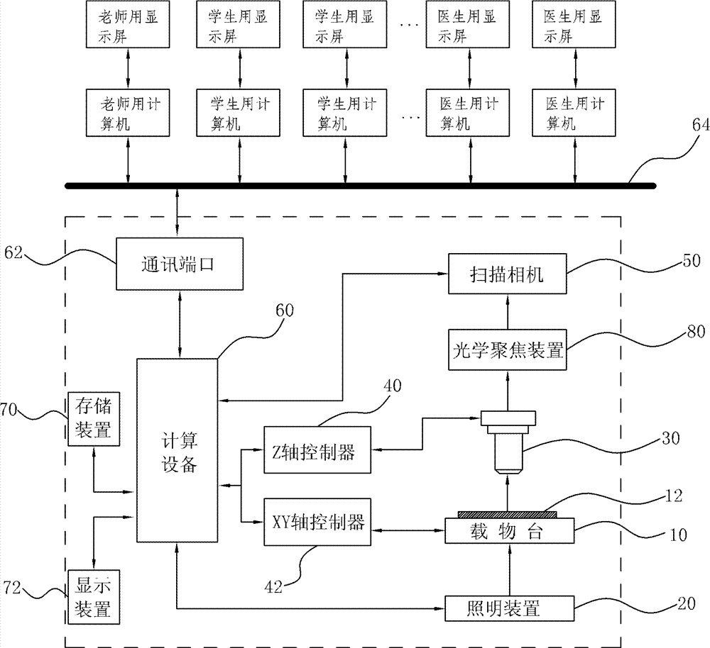
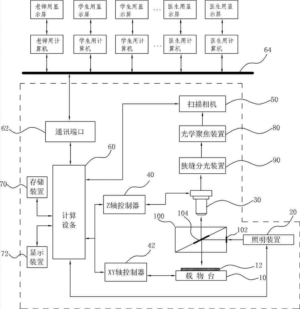
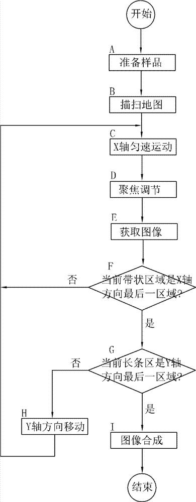

Image

  • Full-automatic scanning system and method for microscopic section
  • Full-automatic scanning system and method for microscopic section
  • Full-automatic scanning system and method for microscopic section

Examples


Embodiment Construction

[0031] In order to make the technical problems, technical solutions and beneficial effects to be solved by the present invention clearer and clearer, the present invention will be further described in detail below in conjunction with the accompanying drawings and embodiments. It should be understood that the specific embodiments described here are only used to explain the present invention, not to limit the present invention.

[0032] Such as figure 1 As shown, the fully automatic scanning system for microsections of the present invention includes an object stage 10 , an illuminating device 20 , an objective lens 30 , and a Z-axis controller 40 . The stage 10 is used to place the sliced ​​sample 12, and drives the sliced ​​sample 12 to move at a uniform speed along the X-axis direction and the Y-axis. The scanning camera 50 is placed at the detector position of the traditional digital microscopic imaging system, but the camera 50 needs to be The imaging surface of the scannin...


PUM


Abstract

The invention discloses a full-automatic scanning system and a method for a microscopic section. The system comprises an object stage, a lighting device, an object lens, a Z-axis controller, a scanning camera and a computing device, the scanning camera is provided with a rolling shutter exposure mode and a banded array or banded subarray reading mode, the scanning camera performs a continuous digital imaging scanning for a banded partial area of a section sample which moves at a constant speed along a shutter rolling direction of the scanning camera, under the rolling shutter exposure mode, the scanning camera can perform a parallel exposure for next frame of image before the exposure of a former frame of image is finished, and the computing device can splice each frame of image which is subjected to continuous exposure to form a complete digital section. By means of the technical scheme, the uniform motion speed of the section sample can be improved, simultaneously the motion blurring caused by sample motion during exposure of the scanning camera can be reduced, the image acquisition speed is improved, and the image acquired by the scanning camera is clear and stable.

Description

technical field [0001] The invention relates to a system and method for scanning microsections, in particular to an automatic scanning system and method for microsections or fluorescent staining microsections. Background technique [0002] In teaching, remote pathological diagnosis, software-based intelligent automatic identification system and other applications, it is necessary to provide the whole picture of the tissue sample observed under the microscope for multiple people to read or to read in different places or at different times without actual section / without microscope read. These applications all require the digitization of the entire or substantial portion of a tissue sample. [0003] When the operator actually observes the tissue sample under the microscope, he generally scans the tissue sample quickly at a low magnification, finds the interesting or suspicious area, and then switches to a high-magnification objective lens to observe the sample partially. Howe...

Claims


Application Information

Patent Timeline
Patent Type & Authority Applications(China)
IPC IPC(8): G02B21/00G02B21/36H04N1/047
Inventor 易定容
Owner MOTIC CHINA GRP CO LTD
Learn More