Method and device for visualizing the registration quality of medical image datasets

A medical image and data group technology, applied in medical image data management, medical image, image data processing, etc., can solve problems such as misjudgment, achieve simple implementation and save time

Inactive Publication Date: 2013-05-15
SIEMENS AG
View PDF4 Cites 14 Cited by
  • Summary
  • Abstract
  • Description
  • Claims
  • Application Information

AI Technical Summary

Problems solved by technology

A problem that may be encountered when using a non-strict registration algorithm is that it is difficult for an observer of the registration result, i.e., the fused image, to determine which parts of an image have more "elastic" transformations and which parts slightly worse
If, for example, a particularly diagnostically important region of an image is altered by a non-strict registration algorithm, this could lead to far-reaching misjudgments

Method used


Image

  • Method and device for visualizing the registration quality of medical image datasets
  • Method and device for visualizing the registration quality of medical image datasets
  • Method and device for visualizing the registration quality of medical image datasets

Examples


Embodiment Construction

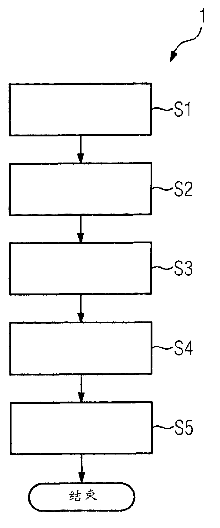
[0035] figure 1 Shown is a flow chart of the method 1 according to the invention for visualizing the registration quality of medical image datasets. Method 1 includes method steps S1-S5, and ends with "END" after method step 5 ends. The following is a detailed description of the method steps:

[0036] S1) collecting a reference image and a target image;

[0037] S2) registering the reference image and the target image into a fused image through a registration method including an inelastic registration method part and an elastic registration method part;

[0038] S3) Determining a deformation region, wherein the deformation region includes a movement vector from an image point of the target image to an image point of the reference image, and the movement vector is partially caused by an elastic registration method;

[0039] S4) superimposing at least one region of the fused image with a superposition mask, and the characteristic parameters of the deformed region enter the su...


PUM


Abstract

A method 1 for visualizing a registration quality of medical image datasets is provided. A reference image 10 and an object image 20 are acquired and registered with each other to form a merged image 40 by a registration method having a non-elastic and an elastic registration method section. A deformation field is determined having displacement vectors 45 from image points 25 of the object image 20 to image points 15 of the reference image 10 which are conditioned by the elastic registration method section. One region of the merged image 40 is overlayed with an overlay 50 into which parameters of the deformation field are fed. The region of the merged image 40 and the overlaid overlay 50 is visualized.

Description

technical field [0001] The invention relates to a method for visualizing the registration quality of medical image datasets. In addition, the invention relates to a corresponding device for visualizing the registration quality of a medical image dataset. Background technique [0002] In interventional radiology, in order to evaluate a case, different modalities are often fused, i.e. superimposed, for example, two-dimensional and three-dimensional X-ray images (XA), computed tomography (CT), magnetic resonance tomography (MR), positron emission tomography (PET), single photon emission computed tomography (SPECT) or ultrasonography (US) image data sets. Physicians can thus combine the respective advantages of the different methods when encountering a particular case. In an ideal case, only basic data image processing operations, such as rotation or translation, should be performed in order to superimpose two images, in order to properly overlay the image. In this case, a st...

Claims


Application Information

Patent Timeline
Patent Type & Authority Applications(China)
IPC IPC(8): G06T7/00G06F19/00
CPCA61B8/13G06T2207/30168G06F19/00G06T2207/10072A61B6/03G06F19/3406A61B19/00A61B6/00G06T7/0024G06F19/321G06T2207/30004G06T2207/20016G06T2200/24A61B5/055G06T7/30G16H40/63G16H30/40G06T1/00H04N5/262G06T7/00G16Z99/00G09G5/00
Inventor S·劳滕施拉格M·特里尼
Owner SIEMENS AG
Why Eureka
Try Eureka