Dynamic optical tomographic imaging devices, methods and systems

A technology of optical tomography and imaging systems, applied in the fields of optical devices, measuring devices, medical science, etc., can solve problems such as dependence, insufficient physiological details, and inability to visualize arterial segments

Active Publication Date: 2013-10-09
THE TRUSTEES OF COLUMBIA UNIV IN THE CITY OF NEW YORK
View PDF5 Cites 10 Cited by
  • Summary
  • Abstract
  • Description
  • Claims
  • Application Information

AI Technical Summary

Problems solved by technology

Duplex ultrasound is limited by its inability to visualize the 13% of the arterial segment at the calf and is dependent on operator technique
[0014]Thus, while X-ray and MRI techniques provide a lot of anatomical detail, they do not provide enough physiological detail, and small arteries are Visualization may not be possible, or not enough blood is flowing through the artery as expected

Method used


Image

  • Dynamic optical tomographic imaging devices, methods and systems
  • Dynamic optical tomographic imaging devices, methods and systems
  • Dynamic optical tomographic imaging devices, methods and systems

Examples


Embodiment Construction

[0031] Embodiments disclosed herein provide a non-invasive optical tomography modality that can be used to detect and monitor peripheral arterial disease (PAD) in the lower extremities. Imaging of peripheral hemodynamics in the lower extremities can effectively diagnose LEAD regardless of arterial compressibility, which is ideal for diabetic patients. Optical tomography can also be used to image vascular dynamics and blood flow patterns. While in breast cancer and arthritis vascular changes are secondary effects, in PAD vascular changes are a primary effect, and the optical tomography modality disclosed in the present invention can be used to diagnose and monitor vascular disease. Breast imaging systems can be used to collect preliminary data in diabetic patients with PAD. In these preliminary images, the difference between healthy people and diabetic PAD patients can be observed.

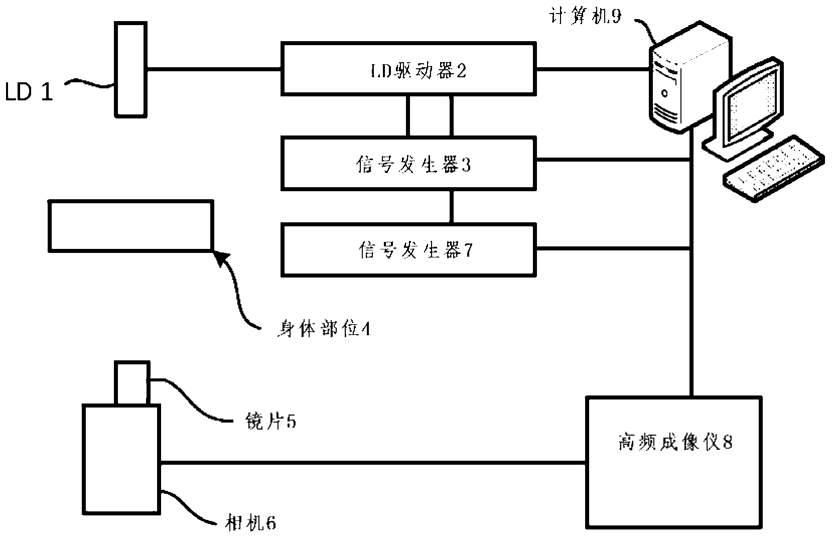
[0032] Figure 1A An optical tomography system 100 is shown for use in one embodiment. Freq...


PUM


Abstract

The disclosed subject matter includes optical tomographic systems for acquiring and displaying dynamic data representing changes in a target tissue sample to external provocation. For example, the disclosed devices, methods and systems may be used for quantifying dynamic vascular changes caused by imposed blood pressure changes for diagnosing peripheral artery disease.

Description

[0001] Cross References to Related Applications [0002] This application is an international application claiming the benefit of US Provisional Patent Application No. 61 / 412,717, filed November 11, 2010, which is hereby incorporated by reference in its entirety. [0003] Statement Regarding Federally Funded Research [0004] This invention was made with government support under NIAMS-2R01-AR046255 sponsored by the National Institute of Arthritis and Musculoskeletal and Skin Diseases. The government has certain rights in this invention. field of invention [0005] The present invention relates to a system, device and method for tomography, in particular to a system, device and method for tomography for diagnosis of peripheral artery disease. Background technique [0006] Optical tomography uses signals produced by amplitude attenuation or phase shifting of scattered light from tissue or samples. Incident light is diffusely reflected by the target and exits the surface....

Claims


Application Information

Patent Timeline
Patent Type & Authority Applications(China)
IPC IPC(8): A61B5/022
CPCA61B5/0053A61B5/0073A61B5/02007A61B5/02A61B5/022G01B9/02A61B5/0066A61B5/0261A61B5/14546A61B5/1455
Inventor 安德烈亚斯·H·希尔舍迈克尔·哈利勒拉杰夫·达亚尔金仁庆金允杰
Owner THE TRUSTEES OF COLUMBIA UNIV IN THE CITY OF NEW YORK
Why Eureka
Try Eureka