Positron emission tomography attenuation correction method and combined tomographic imaging system

A technology of positron emission and tomography, which is applied in computerized tomography scanners, instruments for radiological diagnosis, and measurement using nuclear magnetic resonance imaging systems, and can solve problems such as small MRT data

Active Publication Date: 2016-12-28
SIEMENS HEALTHCARE GMBH
View PDF4 Cites 0 Cited by
  • Summary
  • Abstract
  • Description
  • Claims
  • Application Information

AI Technical Summary

Problems solved by technology

This may not, or only to a lesser extent, provide matched MRT data with no or only small motion differences from the corresponding PET data

Method used


Image

  • Positron emission tomography attenuation correction method and combined tomographic imaging system
  • Positron emission tomography attenuation correction method and combined tomographic imaging system
  • Positron emission tomography attenuation correction method and combined tomographic imaging system

Examples


Embodiment Construction

[0079] Hereinafter, embodiments of the present invention will be described in detail with reference to the accompanying drawings, wherein like reference numerals refer to like elements. It should be understood that the following description of the examples should not be considered limiting. The scope of the present invention is not intended to be limited by the embodiments described below or by the merely illustrative drawings.

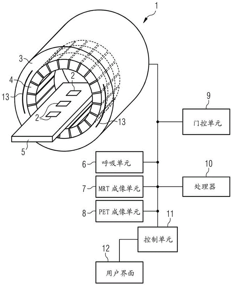
[0080] figure 1 is a schematic diagram of the combined PET-MRT system 1 . A patient can be placed on the table 5 and positioned within the magnet 3 . Magnet 3 can apply a static (DC) magnetic field of several Teslas to align the nuclear spins. The magnet 3 may comprise a superconducting coil in liquid helium.

[0081] A PET imaging unit 8 and a PET detector 4 are provided; they are configured to acquire PET data. The PET detectors measure the coincident PET photons, and the PET imaging unit 8 provides PET data based on these measurements. The de...


PUM


Abstract

The invention relates to an attenuation correction method for positron emission tomography data, and a combined positron emission tomography and magnetic resonance tomography system. Various embodiments relate to methods of attenuation correction of positron emission tomography (PET) data based on magnetic resonance tomography (MRT) data. The method also includes determining other data indicative of a cycle period of the patient's physiological observables, and matching the PET data to the MRT data based on these other data.

Description

technical field [0001] The present invention relates to a method for attenuation correction of positron emission tomography (PET) data based on magnetic resonance tomography (MRT) data acquired from a patient, and a combined PET-MRT system. In particular, various embodiments relate to techniques for matching PET data and MRT data based on other data indicative of a cyclic period of a patient's physiological observables, such as a respiratory cycle or a cardiac cycle. Background technique [0002] In the prior art techniques are known which allow attenuation correction of acquired PET data based on acquired MRT data using combined PET-MRT systems. [0003] As part of the attenuation correction, the attenuation of PET photons emitted due to the interaction of positrons and electrons is determined for their path through absorbing tissue towards the PET detector of the PET imaging unit. The signal detected by the PET imaging unit via the PET detector is corrected in order to el...

Claims


Application Information

Patent Timeline
Patent Type & Authority Patents(China)
IPC IPC(8): A61B6/03A61B5/055G01R33/48
CPCA61B5/055A61B5/0035A61B5/7285A61B6/037A61B6/4417A61B6/5205A61B6/5247A61B6/5258A61B6/5264A61B6/527A61B6/5288A61B6/548G01R33/481
Inventor J.鲍M.芬切尔W.C.豪K.贾特克A.斯坦默
Owner SIEMENS HEALTHCARE GMBH
Why Eureka
Try Eureka