Integrated IVUS (intravascular ultrasound) image and DSA (digital subtraction angiography) image integrating online real-time treatment device

An integrated blood vessel, real-time processing technology, used in catheters, computed tomography scanners, surgery, etc.

Active Publication Date: 2013-12-25
ZHEJIANG UNIV
View PDF4 Cites 2 Cited by
  • Summary
  • Abstract
  • Description
  • Claims
  • Application Information

AI Technical Summary

Problems solved by technology

[0005] For some diffuse vascular lesions, vascular ostia lesions, and in-stent restenosis, which require precise positioning of the stent release point, IVUS guidance is often required. Blind spots, etc., it is difficult to accurately combine IVUS images with DSA images, and it is more difficult to further ensure the quality of percutaneous coronary intervention (PCI)

Method used


Image

  • Integrated IVUS (intravascular ultrasound) image and DSA (digital subtraction angiography) image integrating online real-time treatment device
  • Integrated IVUS (intravascular ultrasound) image and DSA (digital subtraction angiography) image integrating online real-time treatment device

Examples


Embodiment Construction

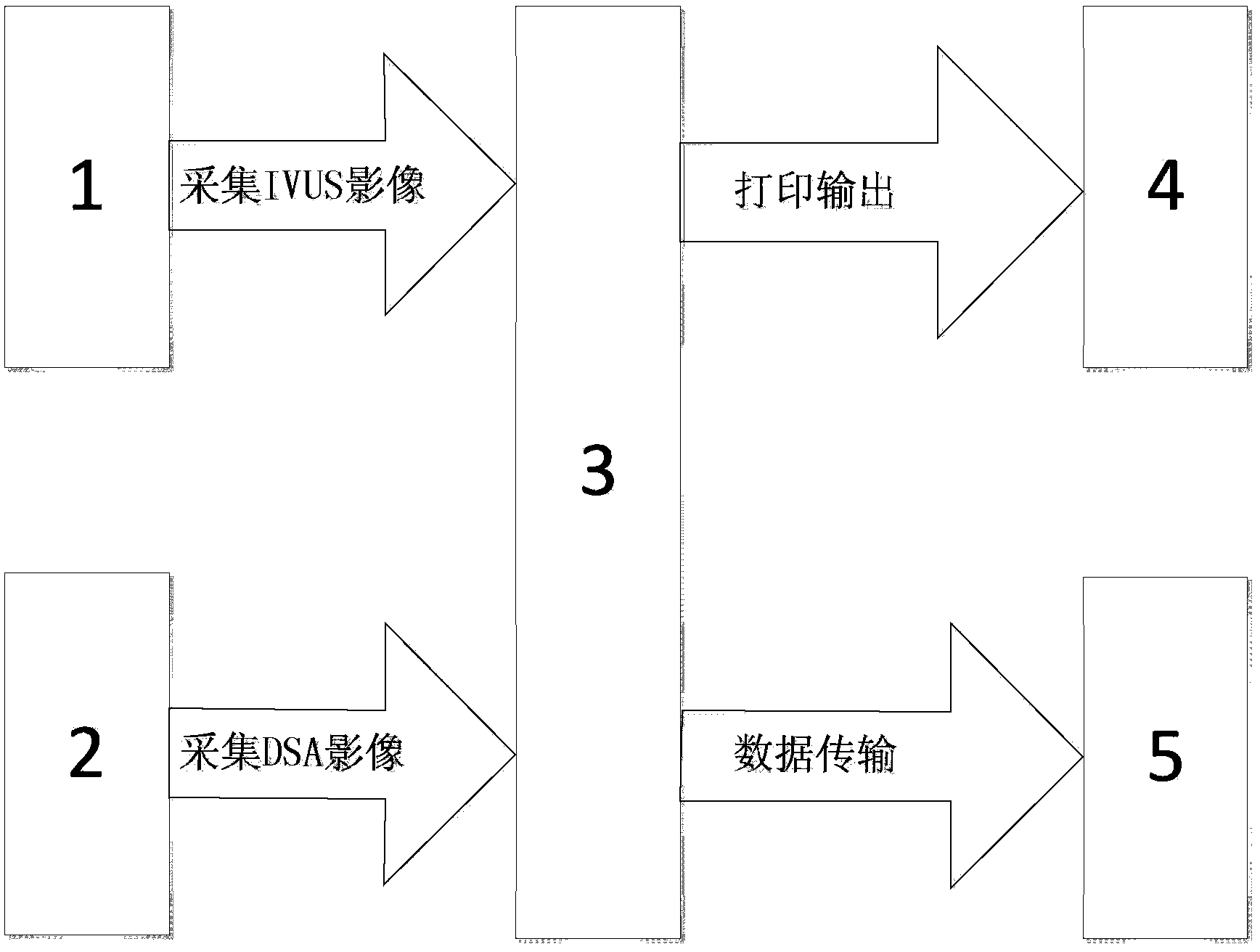
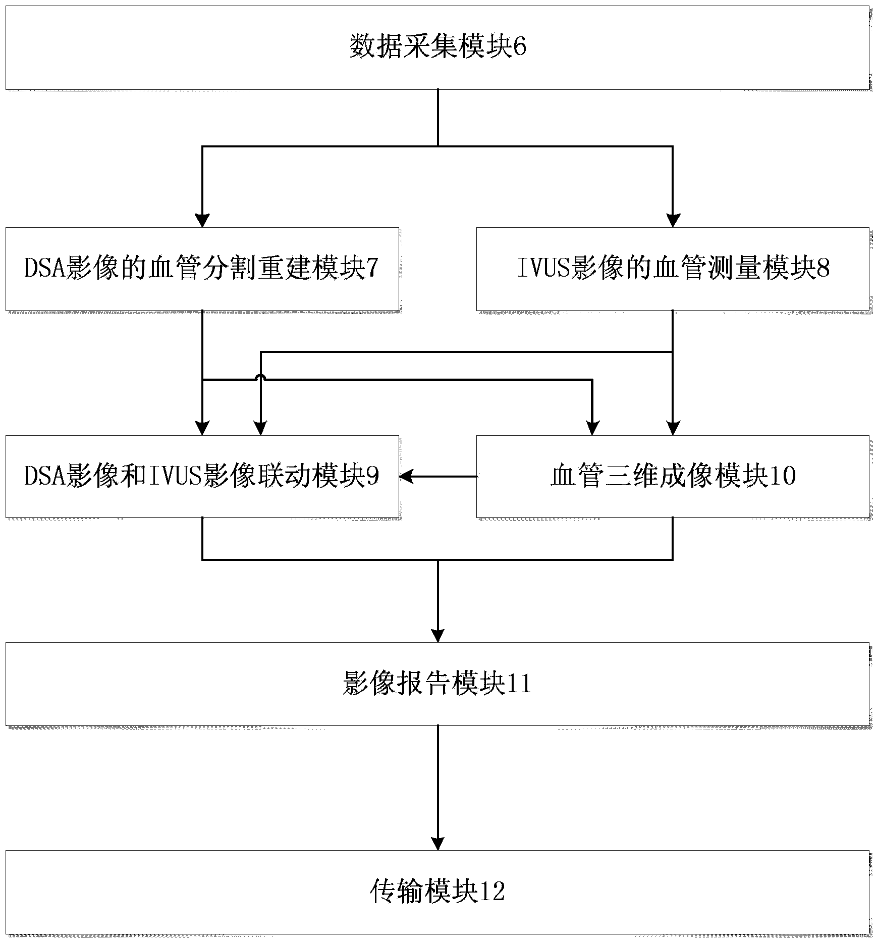
[0018] according to figure 1 and figure 2, the present invention provides an integrated online real-time processor that integrates intravascular ultrasound (IVUS) images and digital subtraction (DSA) images. The system includes seven modules: data acquisition module, vessel segmentation and reconstruction module for DSA images, vessel measurement module for IVUS images, three-dimensional vessel imaging module, linkage module for DSA images and IVUS images, image report module and transmission module. Among them: (1) Data acquisition module: including a high-definition acquisition card with two DVI interfaces embedded in the computer, which is used to collect IVUS images and DSA images in real time during the operation, and store them in the local hard disk of the computer; (2) The blood vessels of DSA images Segmentation and reconstruction module: Based on the matching filtering method, extract the edges, diameters and centerlines of disjoint blood vessels on DSA images, rec...


PUM


Abstract

The invention provides an integrated IVUS (intravascular ultrasound) image and DSA (digital subtraction angiography) image integrating online real-time treatment device, which comprises a data acquisition module, a DSA-image-based blood vessel segmentation and reconstruction module, an IVUS-image-based blood vessel measurement module, a blood vessel three-dimensional imaging module, a DSA image and IVUS image linkage module, an image reporting module and a transmission module. According to the treatment device, a blood vessel is multidimensionally treated by combining a DSA image and an IVUS image; a blood vessel measurement result obtained by the DSA-image-based blood vessel segmentation and reconstruction module is compared with a blood vessel measurement result, obtained by the IVUS-image-based blood vessel measurement module, on a cross section, so that an operator can conveniently judge whether to perform interventional therapy on a blood vessel with lesions or not and determine the position of a blood vessel section to be subjected to the interventional therapy to further determine a therapeutic schedule, and is helped to accurately select the size of a stent and locate the stent.

Description

technical field [0001] The invention belongs to the field of medical equipment, and specifically refers to an integrated online real-time processor integrating intravascular ultrasound (IVUS) images and digital subtraction (DSA) images. Background technique [0002] Coronary angiography is an effective method for the diagnosis of coronary heart disease. Insert the catheter through the femoral artery, radial artery or other peripheral arteries, send it to the ascending aorta, then explore the left or right coronary artery orifice, inject contrast agent, and visualize the coronary arteries, which can clearly reveal the anatomical abnormalities of the coronary arteries The location, degree and scope of its obstructive lesions. Coronary angiography is currently an effective method for directly observing the shape of coronary arteries, and the medical profession calls it the "gold standard". Interventional treatment is required for areas with severe stenosis on angiography. ...

Claims


Application Information

Patent Timeline
Patent Type & Authority Applications(China)
IPC IPC(8): A61B8/12A61B6/03
Inventor 王建安孙勇朱国忠
Owner ZHEJIANG UNIV
Why Eureka
Try Eureka